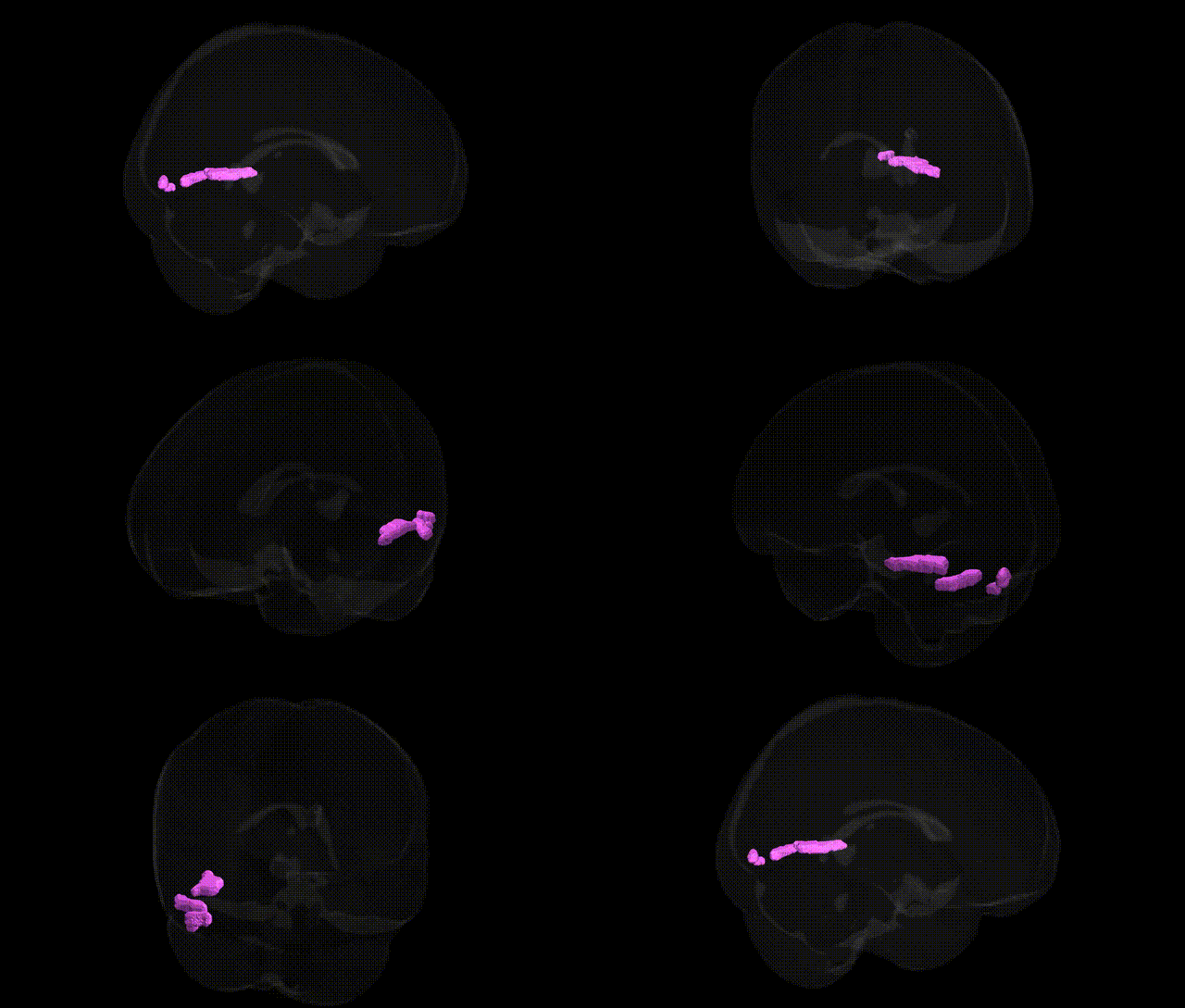
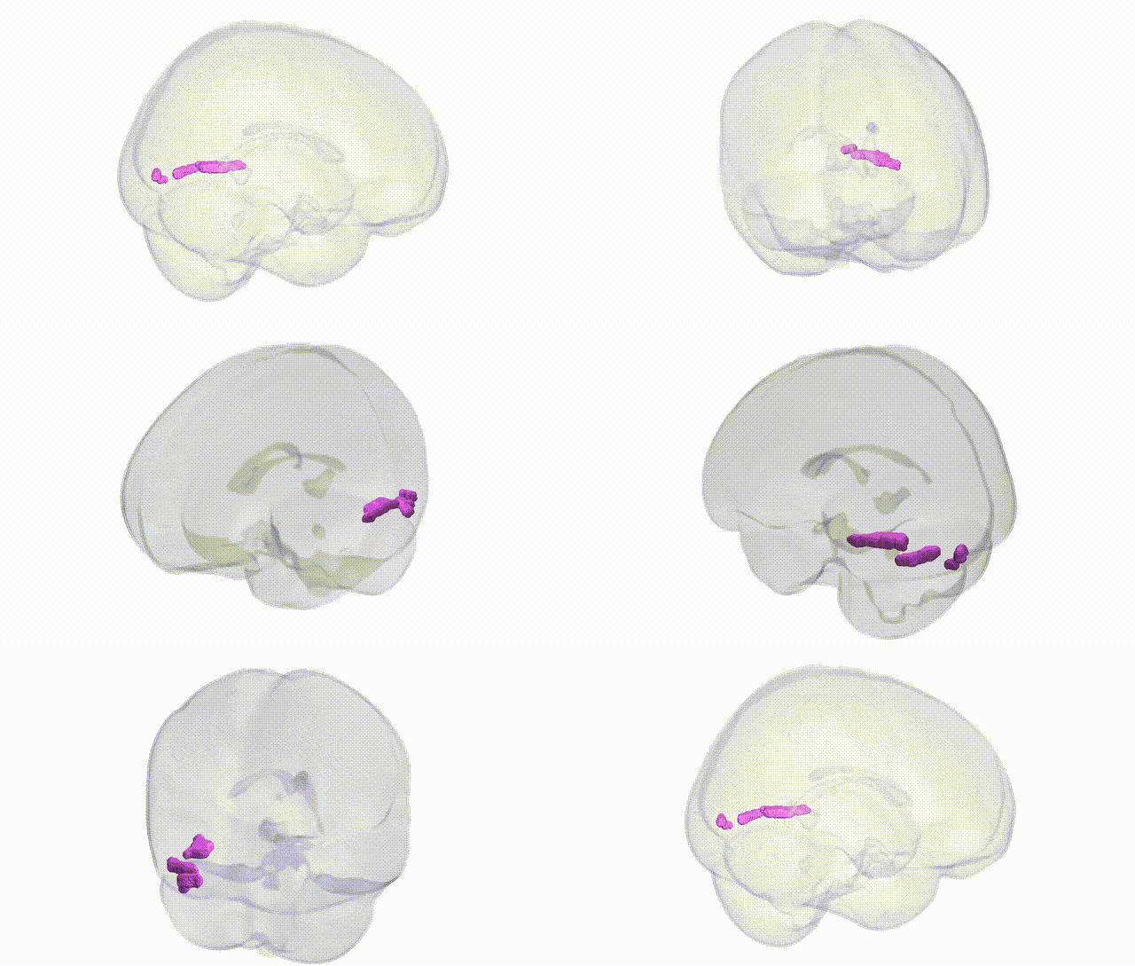
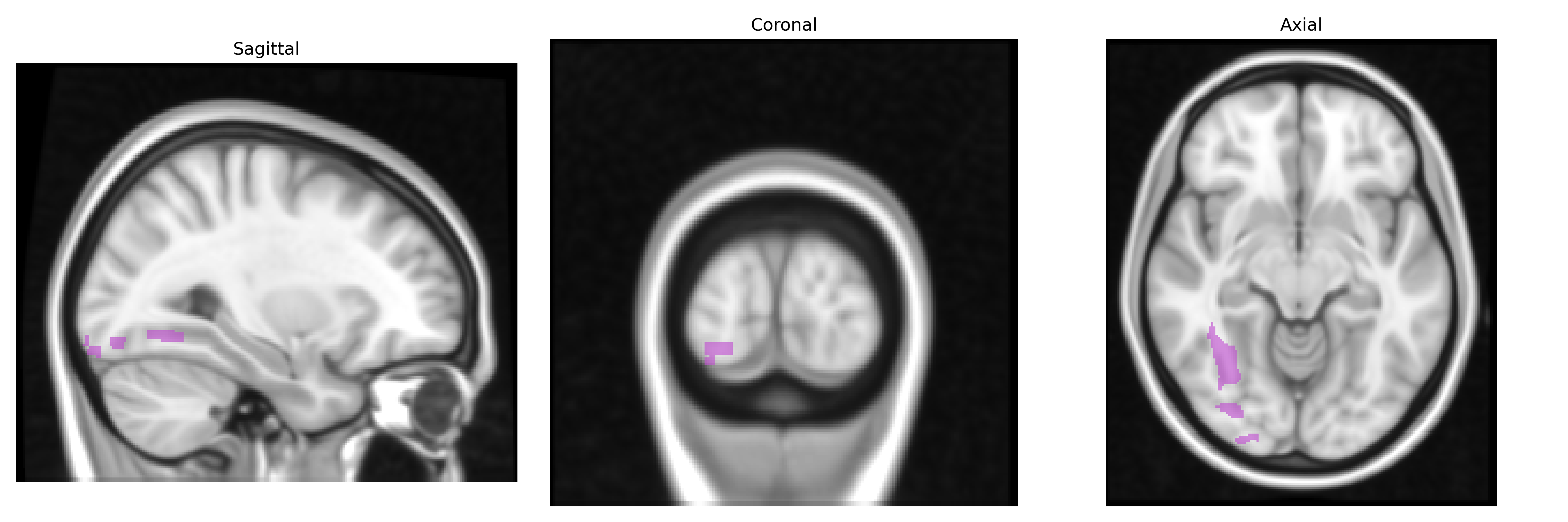
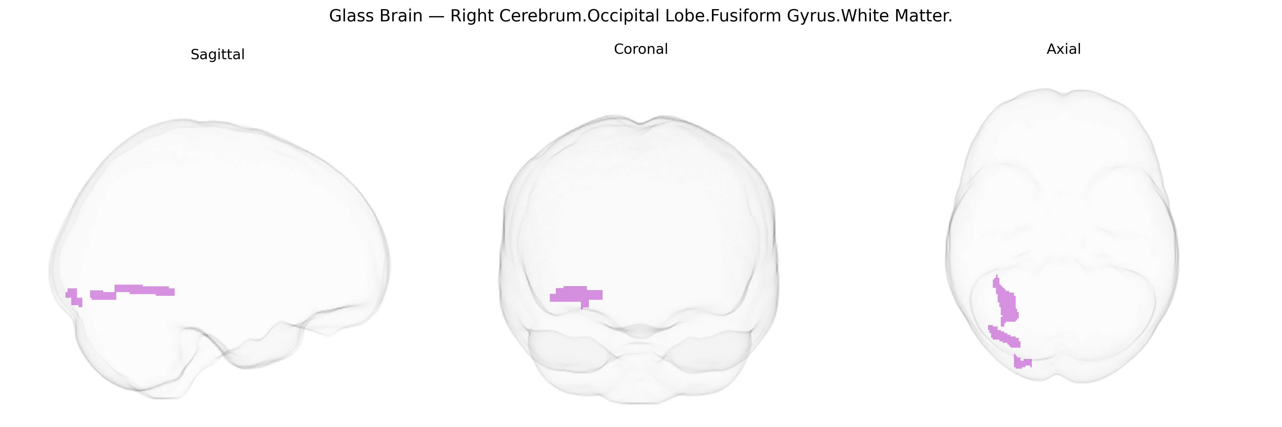
The bilateral Right Cerebrum.Occipital Lobe.Fusiform Gyrus.White Matter corresponds to the deep myelinated fiber pathways underlying the fusiform gyrus in the occipitotemporal region, which connect visual association cortices with temporal and limbic structures. These white matter tracts support high-level visual processing, including object, face, and word recognition, by integrating information between occipital visual areas and anterior temporal regions involved in semantic and memory functions. The fusiform gyrus white matter contains portions of major association pathways such as the inferior longitudinal fasciculus and may interface with the inferior fronto-occipital fasciculus, forming critical conduits for ventral visual stream communication. There is no direct link for this specific white matter label; see the related cortical structure Fusiform gyrus.
The bilateral right occipital fusiform white matter, as defined in the Talairach 1 mm atlas, corresponds largely to occipito-temporal pathways supporting object, face, and word-form processing, and genetic associations in this area are typically inferred from imaging-genetics and GWAS of fusiform/occipital structure and connectivity rather than this exact white-matter label. Polygenic influences on global and regional white matter microstructure (e.g., variants in genes affecting myelination, axonal guidance, and neurodevelopment such as BDNF, NTRK1/2, and multiple oligodendrocyte-related loci) have been linked to diffusion metrics and volume in occipital and fusiform-adjacent tracts in large consortia studies (e.g., ENIGMA, UK Biobank). GWAS of cortical surface area and thickness have repeatedly identified common variants near genes involved in neuronal proliferation (e.g., HMGA2, IGF1-related pathways, and Wnt signaling components) that partly explain inter-individual differences in occipital and fusiform morphology, while studies of the visual word form area and fusiform face area implicate polygenic overlap with cognitive traits such as reading ability, face recognition, and general intelligence. Genetic risk for neurodevelopmental and psychiatric disorders—including autism spectrum disorder, schizophrenia, and major depression—has been associated with altered fusiform and occipital white-matter integrity, with polygenic risk scores for these conditions correlating with microstructural changes in occipito-temporal tracts; in autism, for example, variants influencing synaptic genes (e.g., in the neurexin/neuroligin and SHANK families) and chromatin regulators have been linked to abnormalities in fusiform connectivity relevant to social perception. Additional associations include migraine and visual aura (with multiple GWAS loci near ion-channel and vascular genes showing effects on occipital networks), and rare monogenic leukoencephalopathies (e.g., due to PLP1, ARSA, or other myelin-related genes) that can involve occipital-fusiform white matter, though these are not specific to the right hemisphere. Overall, current genetic findings indicate that variability in this region’s white matter reflects a distributed polygenic architecture shared with broader occipito-temporal and association networks rather than region-specific loci, and is mechanistically tied to genes governing neurodevelopment, myelination, and synaptic function.
Overview generated by GPT-4o (2026).
Region ID: 207
Hemisphere: bilateral
Atlas: Talairach labels 1mm
Full Quality Version: Download MP4
Full Quality Version: Download MP4
Wali Sidiqyar*, Gaurav Rudravaram*, Elyssa M. McMaster, Trent M. Schwartz, Adam M. Saunders, Kurt G. Schilling, Bennett A. Landman "Introducing SPINS: A Shared Public Visualization Library of Neuroanatomical Structures." Medical Imaging with Deep Learning- short paper
This resource is licensed under CC0 1.0 Universal (Public Domain).