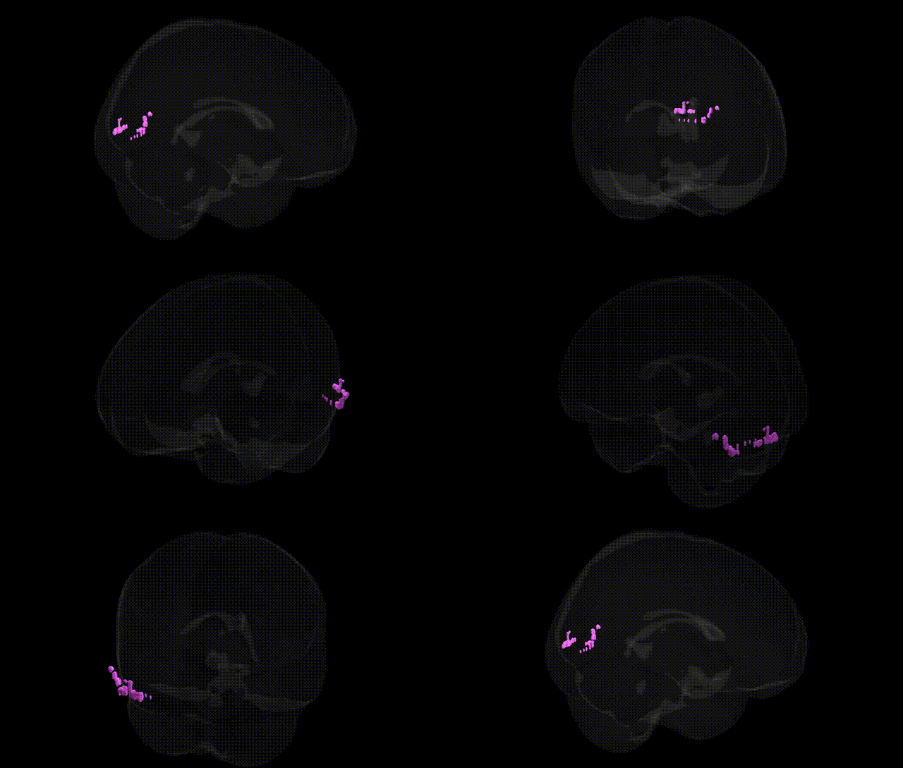
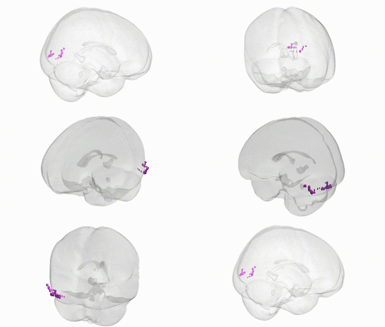
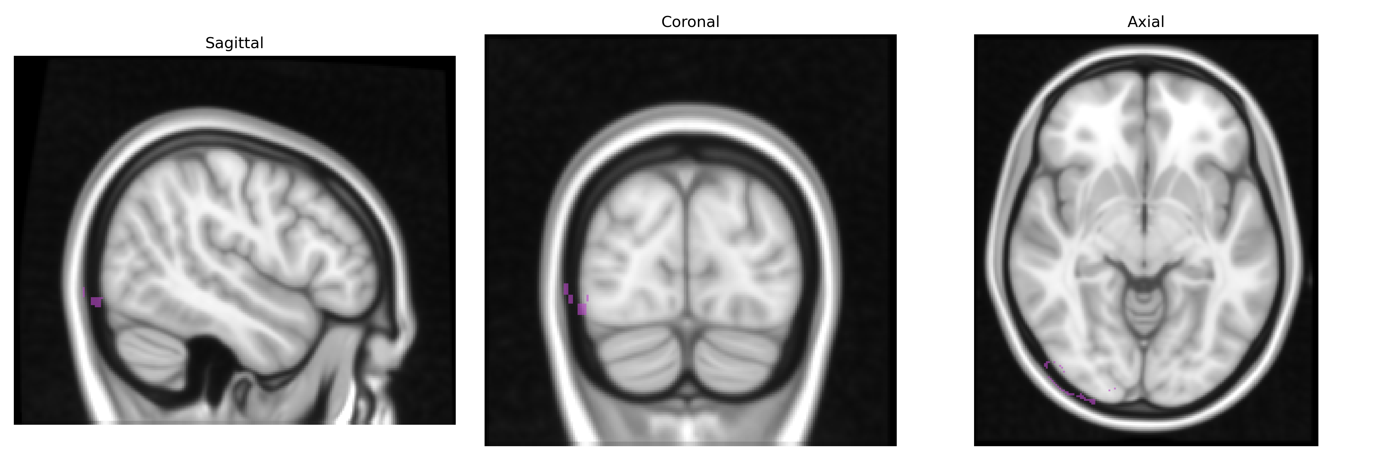
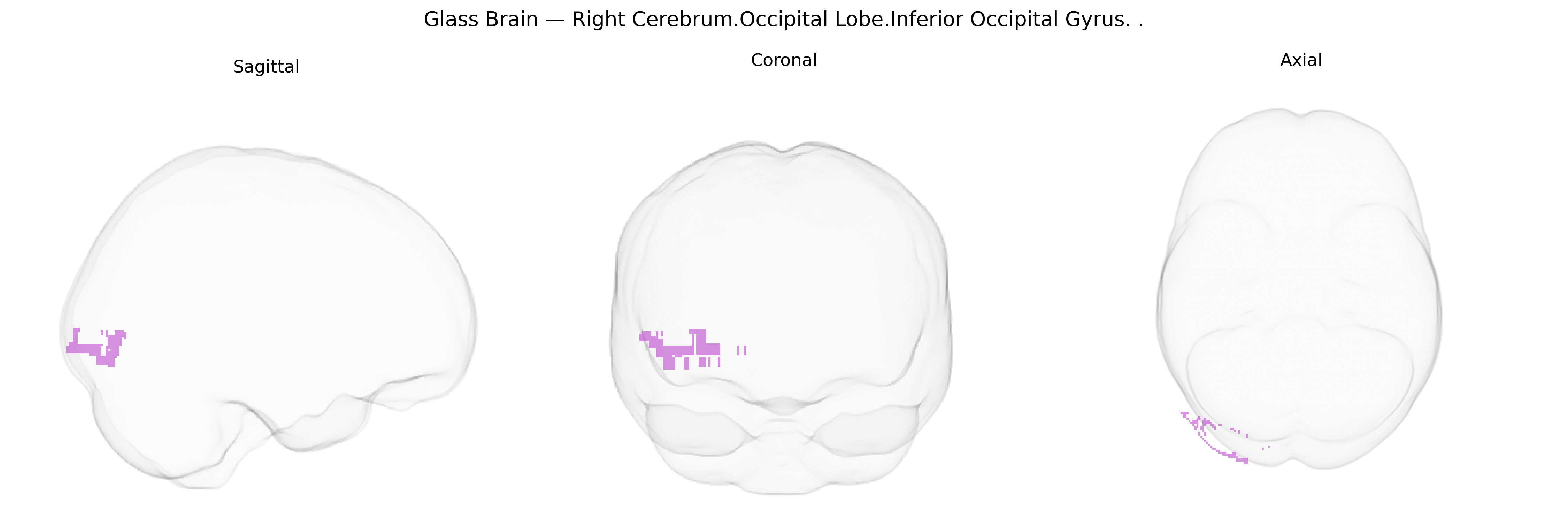
The bilateral Right Cerebrum.Occipital Lobe.Inferior Occipital Gyrus corresponds to cortical tissue located in the inferior portion of the occipital lobe, adjacent to the temporal–occipital junction and ventral visual areas. This gyrus forms part of the visual association cortex involved in higher-order processing of visual input, including aspects of object recognition and integration of complex visual features, and it is closely related to regions such as the lateral occipital complex and portions of the ventral visual stream. Cytoarchitectonically, it lies within extrastriate cortex and receives input from primary visual areas, contributing to the transformation of basic visual signals into more elaborate perceptual representations. There is no direct link for the Inferior Occipital Gyrus; a related structure is the occipital lobe: Occipital Lobe.
Genetic associations specifically targeting the bilateral right inferior occipital gyrus (as defined in the Talairach 1 mm atlas) remain sparse, but converging evidence from imaging genetics and GWAS implicates this region and adjacent occipital areas in several heritable traits and disorders centered on visual processing. Polygenic influences on occipital cortical thickness and surface area have been reported in large-scale MRI–GWAS consortia (e.g., ENIGMA), with loci near genes such as HMGA2, MIR2113, and IGF1 associated with global and regional occipital morphology and visual cortex volume, suggesting broad neurodevelopmental control of occipital structure. Functional imaging genetics studies indicate that variants in genes related to synaptic plasticity and neurotransmission (for example, BDNF Val66Met, COMT Val158Met, and dopaminergic pathway genes) can modulate activation in inferior occipital regions during face and object recognition, consistent with the role of this gyrus in early visual and category-specific processing. Candidate gene and GWAS-based endophenotype work in autism spectrum disorder, schizophrenia, and major depression has linked polygenic risk scores for these conditions to altered occipital activation or thickness, including the inferior occipital gyrus, though effects are often small and distributed. In developmental dyslexia and other reading-related disorders, genetic variants affecting dorsal-ventral visual pathways (e.g., in KIAA0319 and DCDC2) have been associated with altered activation in occipito-temporal regions that encompass or border the inferior occipital gyrus, particularly within the visual word form system. Migraine and visual aura GWAS highlight loci influencing cortical excitability and vascular regulation (such as near TRPM8 and PRDM16), and imaging correlates sometimes show altered occipital structure and function, implying indirect genetic effects on this region. Overall, current evidence points to polygenic, pleiotropic influences on inferior occipital gyrus structure and function, with genetic effects typically operating at the level of broader occipital or ventral visual networks rather than this gyrus in isolation.
Overview generated by GPT-4o (2026).
Region ID: 257
Hemisphere: bilateral
Atlas: Talairach labels 1mm
Full Quality Version: Download MP4
Full Quality Version: Download MP4
Wali Sidiqyar*, Gaurav Rudravaram*, Elyssa M. McMaster, Trent M. Schwartz, Adam M. Saunders, Kurt G. Schilling, Bennett A. Landman "Introducing SPINS: A Shared Public Visualization Library of Neuroanatomical Structures." Medical Imaging with Deep Learning- short paper
This resource is licensed under CC0 1.0 Universal (Public Domain).