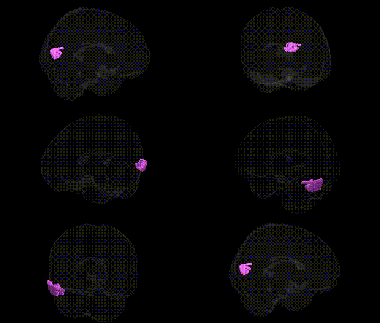
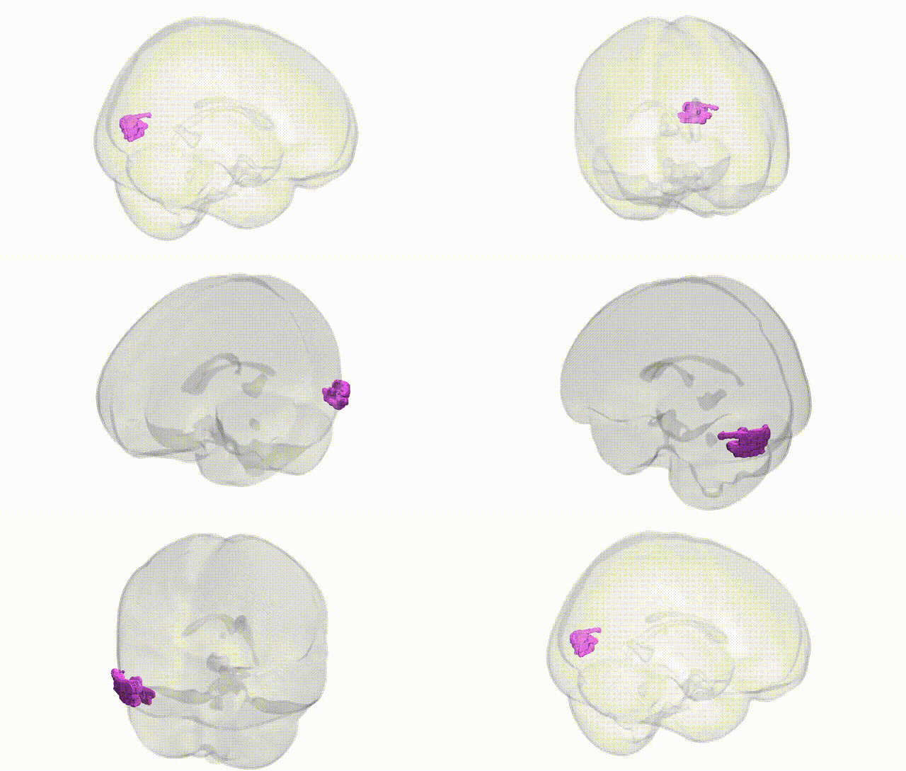
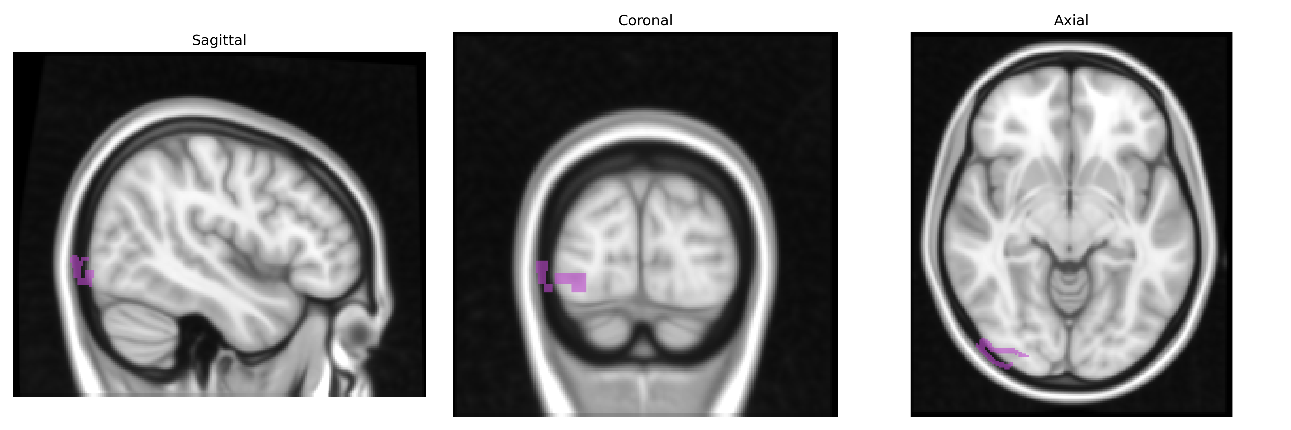
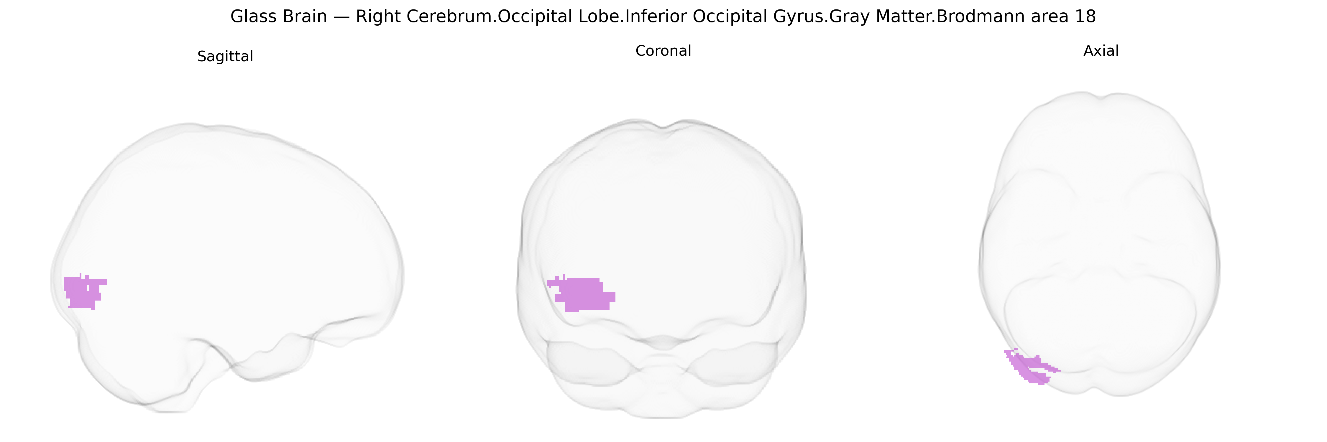
The bilateral Right Cerebrum.Occipital Lobe.Inferior Occipital Gyrus.Gray Matter.Brodmann area 18 corresponds to a portion of the secondary visual cortex (visual association area) located in the caudal occipital lobe, lateral and inferior to the primary visual cortex (BA17). This region participates in early-stage processing of complex visual information, including contours, orientation, and motion-related features, and contributes to the integration of visual signals for object recognition and higher-order visual perception. Neurons in BA18 receive dense feedforward input from BA17 and project to higher-tier visual cortices (such as BA19 and temporal visual areas), forming part of the extrastriate visual network that supports the transformation of basic retinal inputs into increasingly abstract visual representations. There is no direct Wikipedia article for “Inferior Occipital Gyrus BA18”; a related structure is Brodmann area 18.
The bilateral right occipital lobe inferior occipital gyrus gray matter within Brodmann area 18 (secondary visual cortex, V2) has been implicated in several genetic and GWAS-based associations, primarily through imaging-genetics and neuropsychiatric studies. Twin and SNP-based heritability analyses indicate moderate-to-high heritability of cortical thickness and surface area in BA18, with loci including variants near genes involved in neurodevelopment (e.g., MIR137, TBR1, and EMX2-related pathways) and synaptic signaling, as reported in large-scale ENIGMA and UK Biobank imaging GWAS. Visual cortex volume and surface traits encompassing BA18 show associations with polygenic scores for intelligence, educational attainment, and general cognitive ability, consistent with this region’s role in visuospatial and reading-related processing. GWAS of psychiatric disorders (notably schizophrenia, bipolar disorder, and major depression) and autism spectrum disorder have identified risk variants whose carriers display altered occipital cortical thickness or activity in BA18, often interpreted as intermediate endophenotypes rather than direct region-specific genetic effects. Occipital BA18 has also emerged in neurodevelopmental and reading-disorder (dyslexia) imaging-genetic work—implicating genes involved in axon guidance, neuronal migration, and visual word-form processing—and in migraine and visual aura studies, where genetic risk for cortical hyperexcitability is associated with structural and functional differences in early visual cortex. Overall, current evidence reflects polygenic, pleiotropic effects where genetic variants implicated in broad brain development, synaptic plasticity, and psychiatric or cognitive traits contribute to individual differences in BA18 structure and function, rather than a small set of genes uniquely targeting this specific Talairach-defined region.
Overview generated by GPT-4o (2026).
Region ID: 254
Hemisphere: bilateral
Atlas: Talairach labels 1mm
Full Quality Version: Download MP4
Full Quality Version: Download MP4
Wali Sidiqyar*, Gaurav Rudravaram*, Elyssa M. McMaster, Trent M. Schwartz, Adam M. Saunders, Kurt G. Schilling, Bennett A. Landman "Introducing SPINS: A Shared Public Visualization Library of Neuroanatomical Structures." Medical Imaging with Deep Learning- short paper
This resource is licensed under CC0 1.0 Universal (Public Domain).