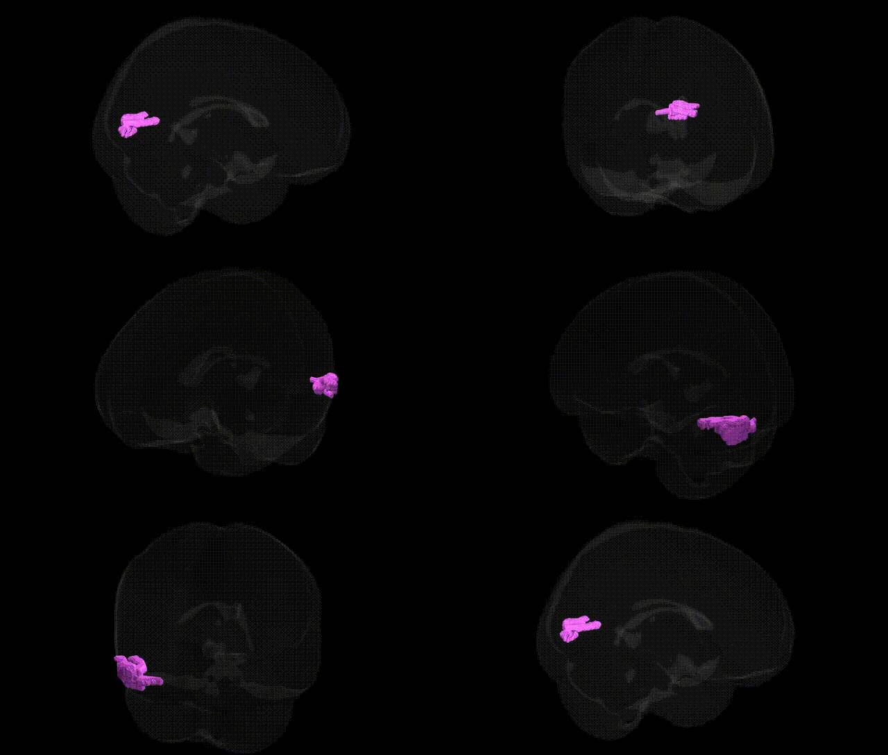
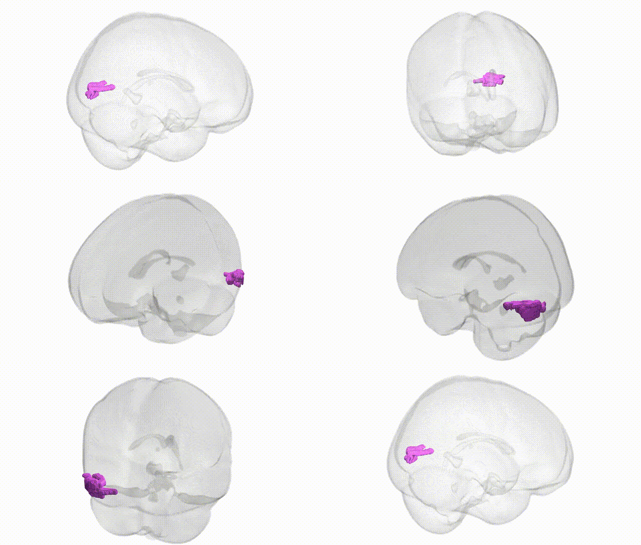
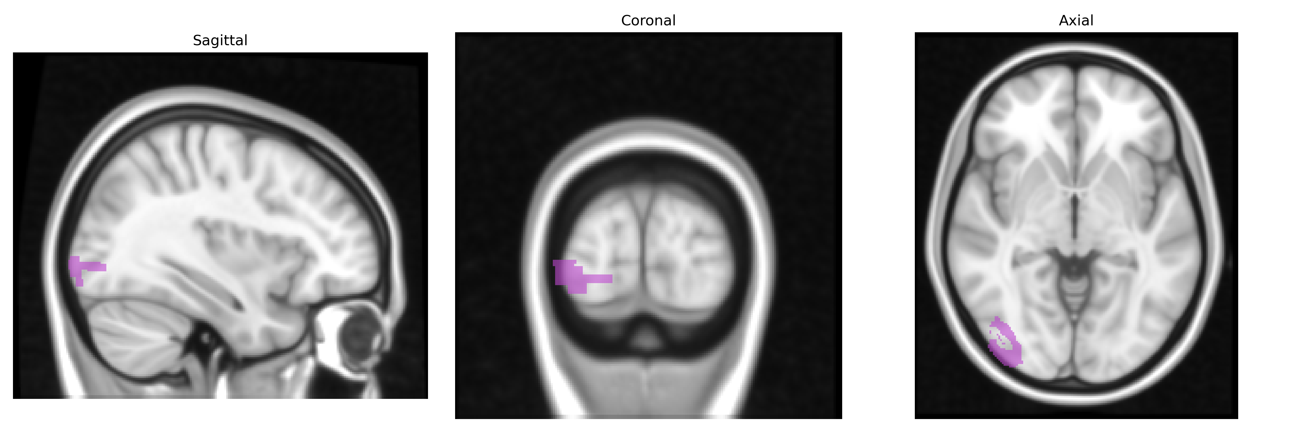
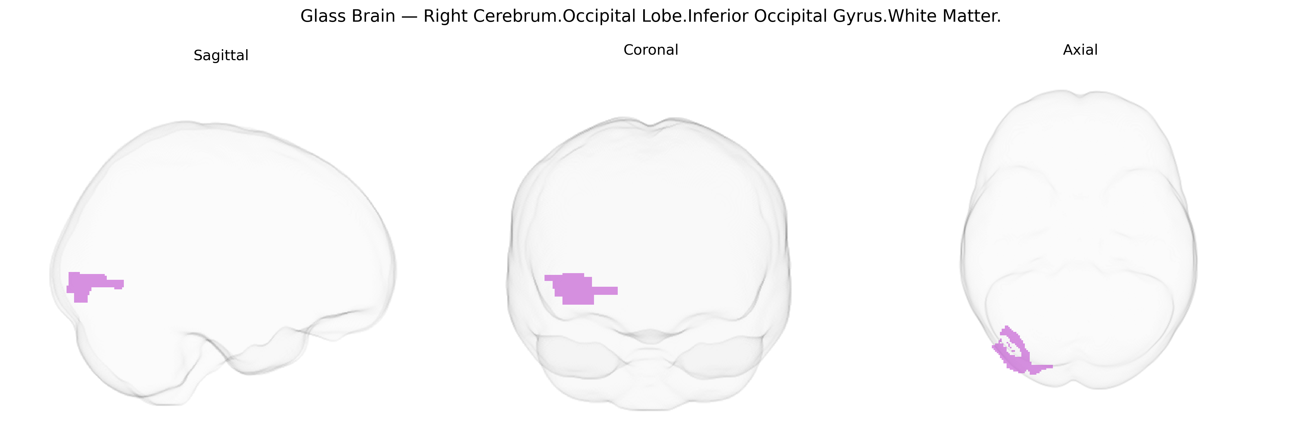
The bilateral right cerebrum occipital lobe inferior occipital gyrus white matter comprises the deep myelinated fiber pathways underlying the inferior occipital gyrus in the right occipital lobe, contributing to the transmission of visual information between primary visual cortex, higher-order ventral visual areas, and more anterior temporal and parietal regions. These fibers include components of long association tracts such as the inferior longitudinal fasciculus and related occipito-temporal connections, which support object recognition, complex form and color processing, and integration of visual input with memory and semantic systems. Damage to this white matter region can disrupt ventral visual stream function, leading to deficits such as visual agnosia or impaired recognition of faces and objects, depending on the extent and connectivity of the lesion. There is no direct link for this exact subregion; a related structure is the Occipital lobe.
The bilateral right inferior occipital gyrus white matter, as defined in the Talairach 1 mm atlas, has not been a primary focus of locus-specific genetic analyses, but converging evidence from imaging genetics and GWAS of occipital and visual-system phenotypes implicates several pathways. Large-scale brain MRI GWAS (e.g., ENIGMA and UK Biobank) have identified common variants in genes involved in axonal guidance, myelination, and synaptic development (such as NRG1, CNTNAP2, and multiple cell-adhesion and oligodendrocyte-related genes) that associate with occipital lobe white matter volume and microstructural measures (e.g., fractional anisotropy) encompassing inferior occipital pathways. Genetic correlations link these occipital white matter traits with neurodevelopmental and psychiatric disorders, including schizophrenia, autism spectrum disorder, and major depression, consistent with observed white matter alterations in these conditions extending into inferior occipital regions. Variants near visual system genes (e.g., those affecting cortical surface area and thickness in visual cortices) have also been associated with global occipital morphology that includes the inferior occipital gyrus territory, and polygenic risk scores for disorders such as schizophrenia and ADHD show subtle associations with occipital white matter integrity in diffusion tensor imaging studies. However, no robust, region-exclusive genetic associations have been reported specifically for the right inferior occipital gyrus white matter, and existing findings primarily reflect broader occipital or visual-network genetic influences rather than a uniquely defined locus.
Overview generated by GPT-4o (2026).
Region ID: 256
Hemisphere: bilateral
Atlas: Talairach labels 1mm
Full Quality Version: Download MP4
Full Quality Version: Download MP4
Wali Sidiqyar*, Gaurav Rudravaram*, Elyssa M. McMaster, Trent M. Schwartz, Adam M. Saunders, Kurt G. Schilling, Bennett A. Landman "Introducing SPINS: A Shared Public Visualization Library of Neuroanatomical Structures." Medical Imaging with Deep Learning- short paper
This resource is licensed under CC0 1.0 Universal (Public Domain).