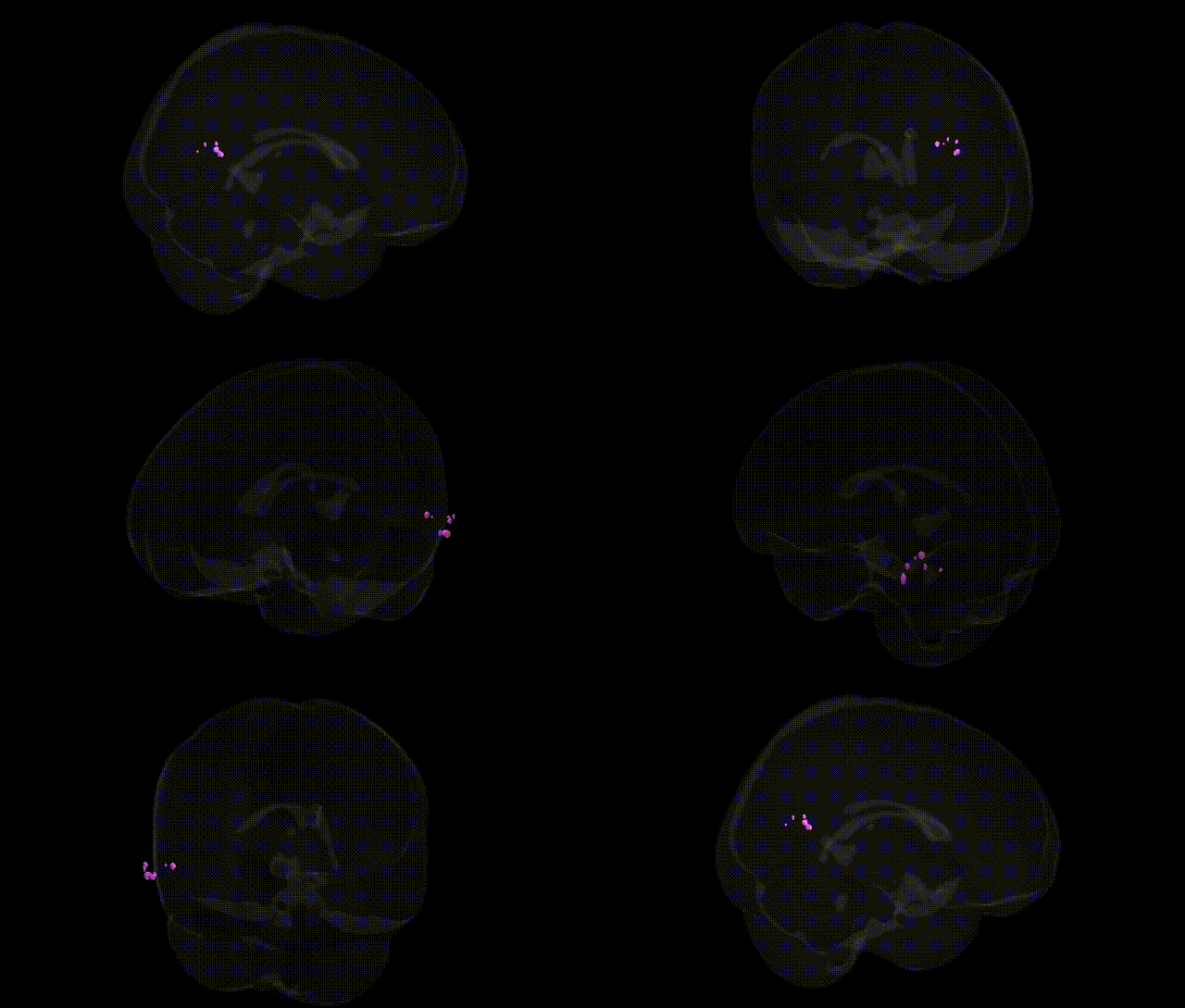
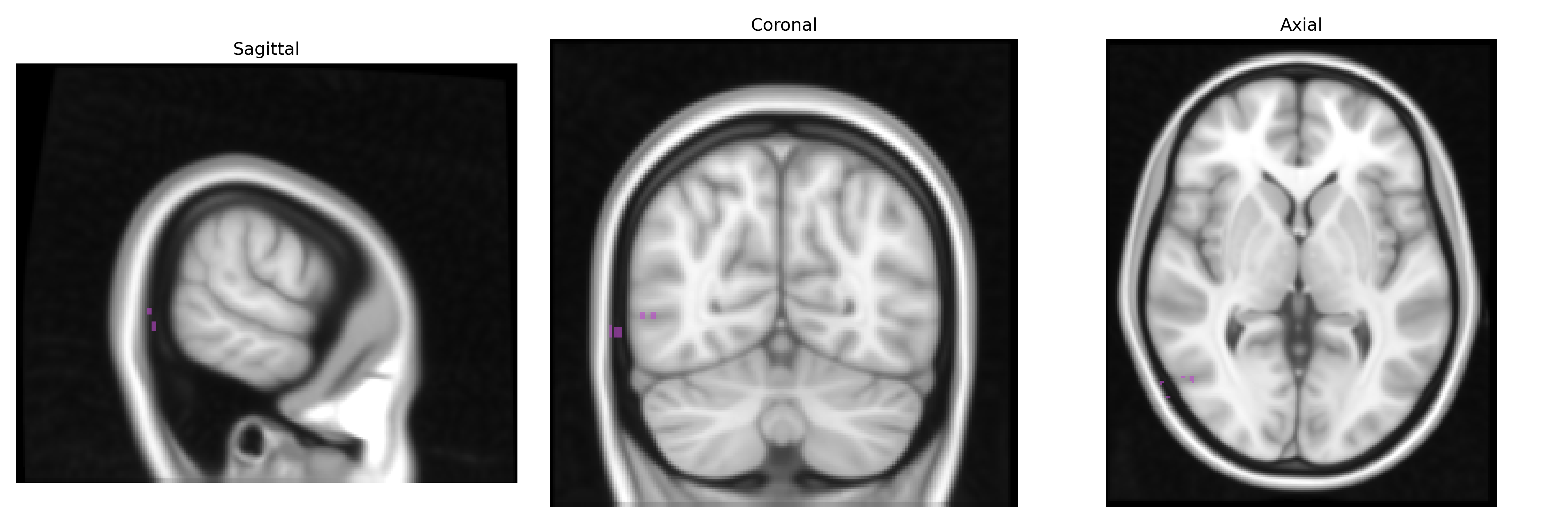
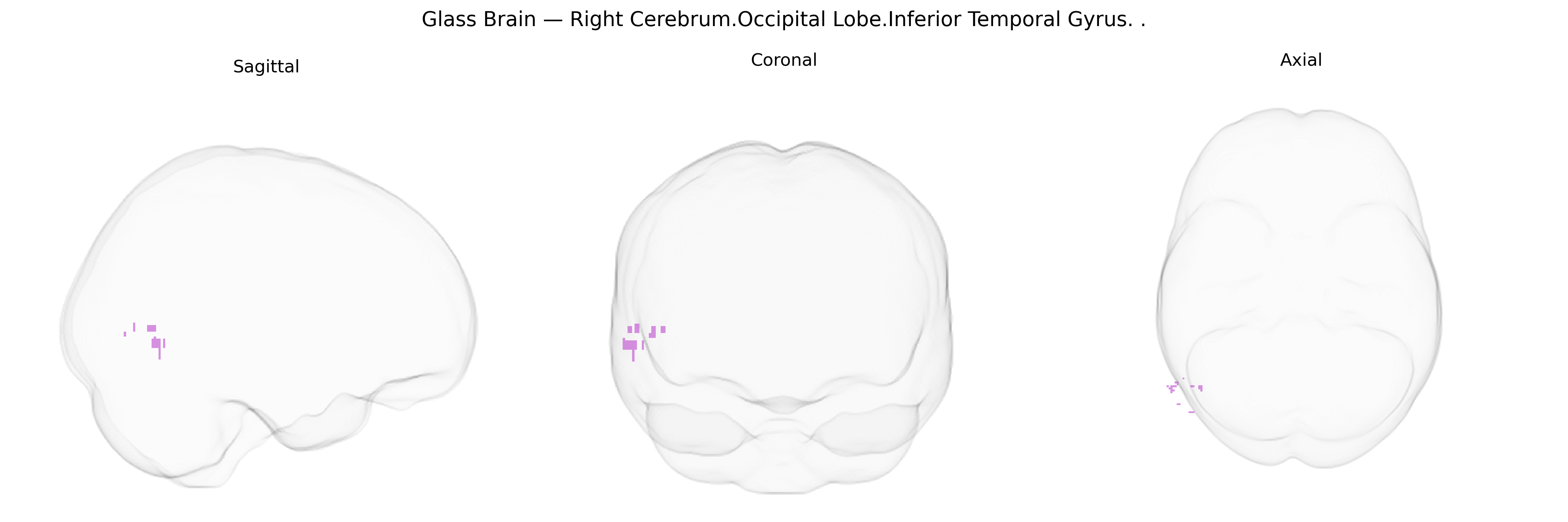
The bilateral Right Cerebrum.Occipital Lobe.Inferior Temporal Gyrus corresponds to the inferior temporal gyrus located near the occipito-temporal junction of the right cerebral hemisphere, extending along the ventral surface of the temporal and occipital lobes. This region forms part of the ventral visual processing stream (“what” pathway), contributing to high-level visual object recognition, including complex shapes, faces, and category-specific visual representations. It receives input from earlier visual areas in the occipital lobe and projects to anterior temporal and other association cortices, supporting visual memory and semantic processing. Functionally, it participates in integrating detailed visual features into coherent percepts and is implicated in disorders such as visual agnosias when damaged. There is no direct link for this exact composite label; a closely related structure is the inferior temporal gyrus: Inferior temporal gyrus.
The bilateral right cerebrum occipital lobe inferior temporal gyrus, corresponding to high-level ventral visual and object-recognition cortex (often overlapping with temporal–occipital regions such as fusiform and inferior temporal areas), has been implicated indirectly in several genetic and GWAS-based findings, although relatively few studies target this exact Talairach-defined parcel. Imaging genetics and large-scale GWAS of cortical thickness, surface area, and regional volumes (e.g., ENIGMA and UK Biobank analyses) have identified multiple loci influencing temporal–occipital morphology, including variants in genes related to neurodevelopment and synaptic function such as KIAA0586, MIR924HG, and others affecting cortical folding and visual association cortex metrics. Polygenic risk for schizophrenia, bipolar disorder, major depression, and autism spectrum disorder has been associated with structural and functional differences in inferior temporal and adjacent fusiform/occipitotemporal regions, consistent with roles in face and object processing and social cognition. GWAS of face recognition ability and prosopagnosia, as well as reading-related traits and dyslexia, have linked risk variants in genes such as DCDC2, KIAA0319, and CNTNAP2 to altered structure or activation of inferior temporal and fusiform regions, though not always isolating the right inferior temporal gyrus specifically. Neurodegenerative and neurodevelopmental disorder genetics (e.g., Alzheimer’s disease risk loci like APOE and CLU, and frontotemporal dementia genes such as MAPT and GRN) also show downstream effects on temporal–occipital cortex atrophy patterns, which often include inferior temporal territory. Overall, genetic associations for this region are largely inferred from broader temporal–occipital or ventral visual stream analyses rather than from studies restricted to the bilateral right inferior temporal gyrus in the Talairach 1 mm atlas.
Overview generated by GPT-4o (2026).
Region ID: 325
Hemisphere: bilateral
Atlas: Talairach labels 1mm
Full Quality Version: Download MP4
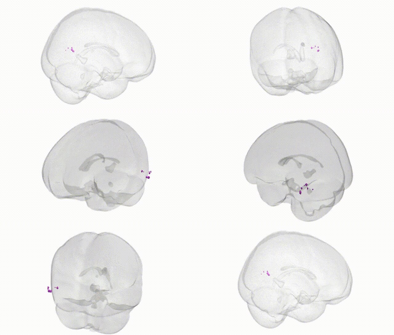
Full Quality Version: Download MP4
Wali Sidiqyar*, Gaurav Rudravaram*, Elyssa M. McMaster, Trent M. Schwartz, Adam M. Saunders, Kurt G. Schilling, Bennett A. Landman "Introducing SPINS: A Shared Public Visualization Library of Neuroanatomical Structures." Medical Imaging with Deep Learning- short paper
This resource is licensed under CC0 1.0 Universal (Public Domain).