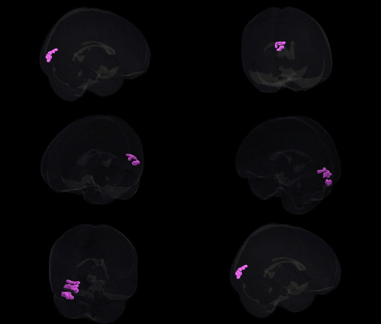
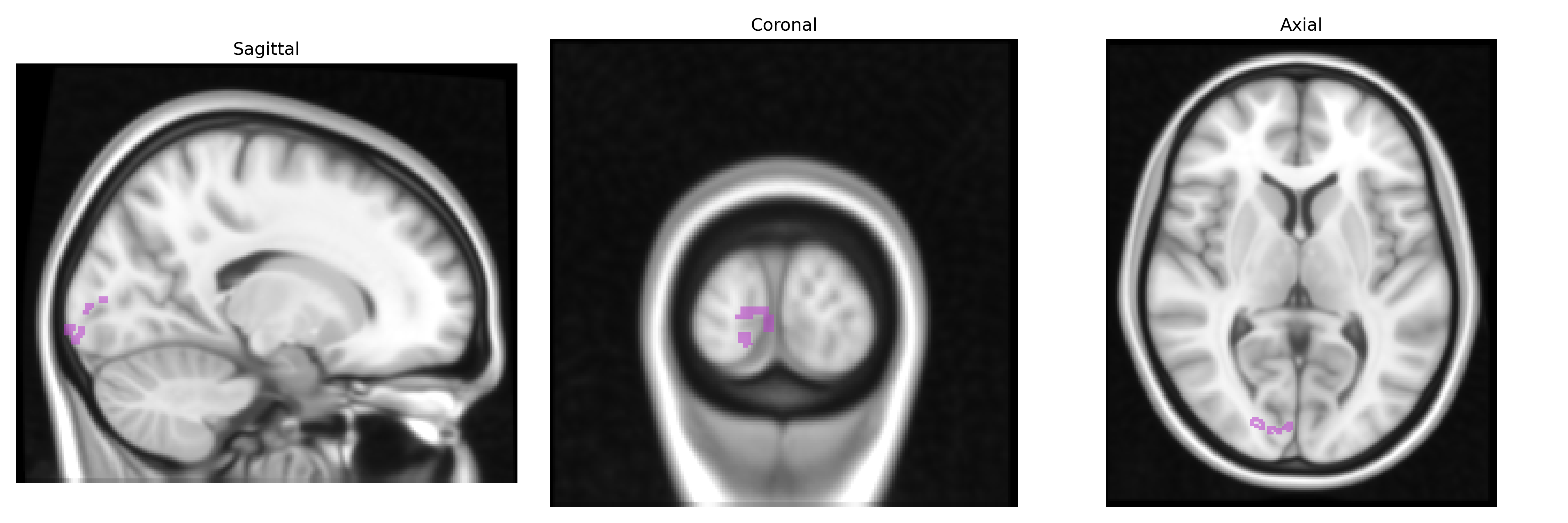
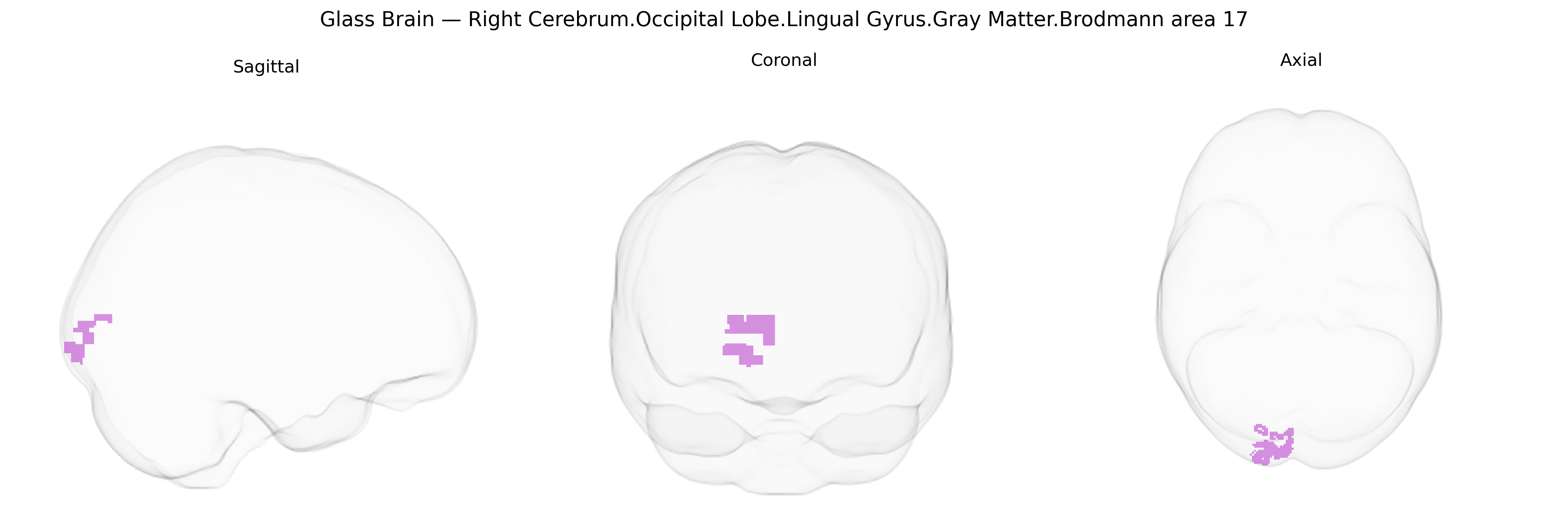
The bilateral Right Cerebrum.Occipital Lobe.Lingual Gyrus.Gray Matter.Brodmann area 17 corresponds to the primary visual cortex (V1) located along the calcarine fissure within the occipital lobe, specifically involving the lingual gyrus on the inferior bank. This region is the main cortical recipient of visual input from the lateral geniculate nucleus of the thalamus and is organized retinotopically, with a precise spatial mapping of the visual field and a disproportionate representation of the central (foveal) visual field. Neurons in this gray matter area exhibit high sensitivity to basic visual features such as orientation, spatial frequency, and contrast, forming the initial stage of cortical visual processing that supports more complex visual perception in downstream extrastriate areas. There is no direct Wikipedia article for this exact Talairach-defined label; a closely related structure is the Brodmann area 17.
The bilateral right lingual gyrus gray matter in Brodmann area 17 (primary visual cortex) has been implicated in multiple genetic and GWAS-based associations, particularly involving visual processing, neurodevelopment, and neuropsychiatric traits. Large-scale imaging–genetics consortia such as ENIGMA and UK Biobank have identified common variants (notably in genes involved in neuronal development, synaptic signaling, and myelination—e.g., PITPNM2, HMGA2, KIAA0586, and loci near MAPT and WNT pathway genes) that influence occipital and specifically lingual gyrus cortical thickness, surface area, and gray matter volume. Polygenic scores for educational attainment, general cognitive ability, and intracranial volume show correlations with structural measures in primary visual cortex, suggesting shared genetic architecture between visual cortical morphology and cognitive traits. Psychiatric GWAS meta-analyses report that risk alleles for schizophrenia, bipolar disorder, and major depression are associated with altered occipital and lingual gyrus structure or activation, consistent with visual and perceptual disturbances in these conditions. Additionally, genetic variants linked to visual acuity, refractive error (e.g., myopia-associated loci near GJD2 and RASGRF1), and migraine (e.g., in TRPM8 and near LRP1) show downstream associations with primary visual cortex structure or function, reflecting the role of this region in early visual processing and susceptibility to visual aura and related phenotypes.
Overview generated by GPT-4o (2026).
Region ID: 245
Hemisphere: bilateral
Atlas: Talairach labels 1mm
Full Quality Version: Download MP4
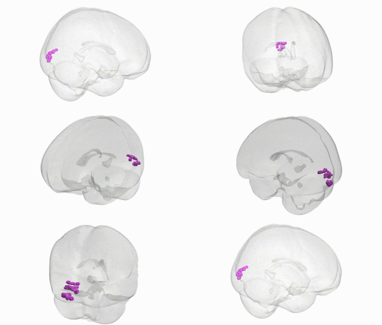
Full Quality Version: Download MP4
Wali Sidiqyar*, Gaurav Rudravaram*, Elyssa M. McMaster, Trent M. Schwartz, Adam M. Saunders, Kurt G. Schilling, Bennett A. Landman "Introducing SPINS: A Shared Public Visualization Library of Neuroanatomical Structures." Medical Imaging with Deep Learning- short paper
This resource is licensed under CC0 1.0 Universal (Public Domain).