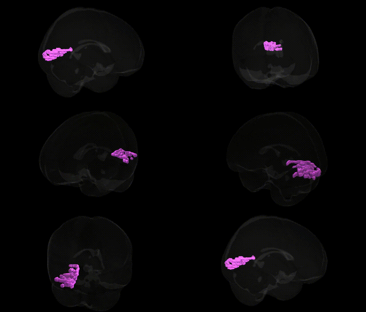
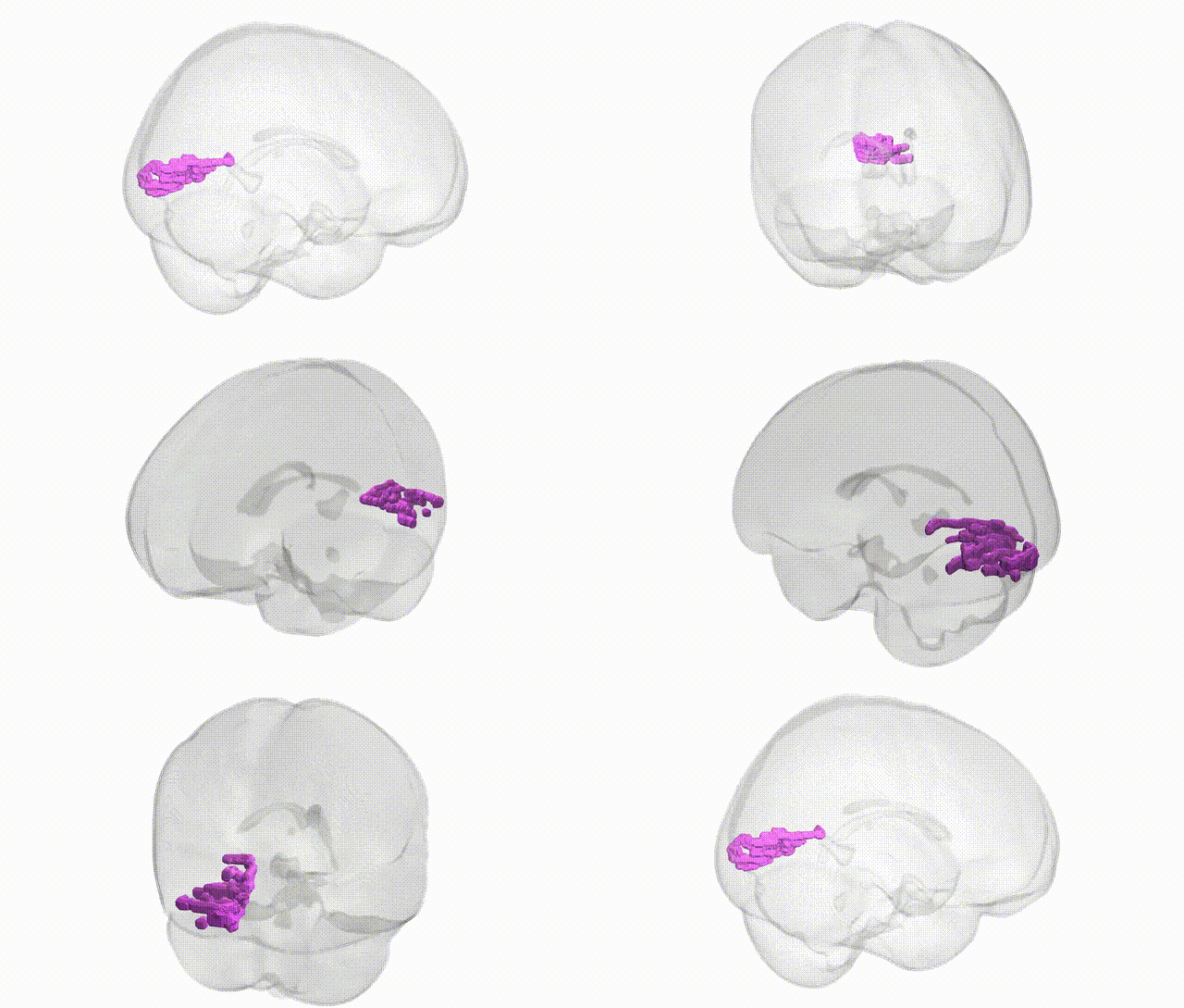
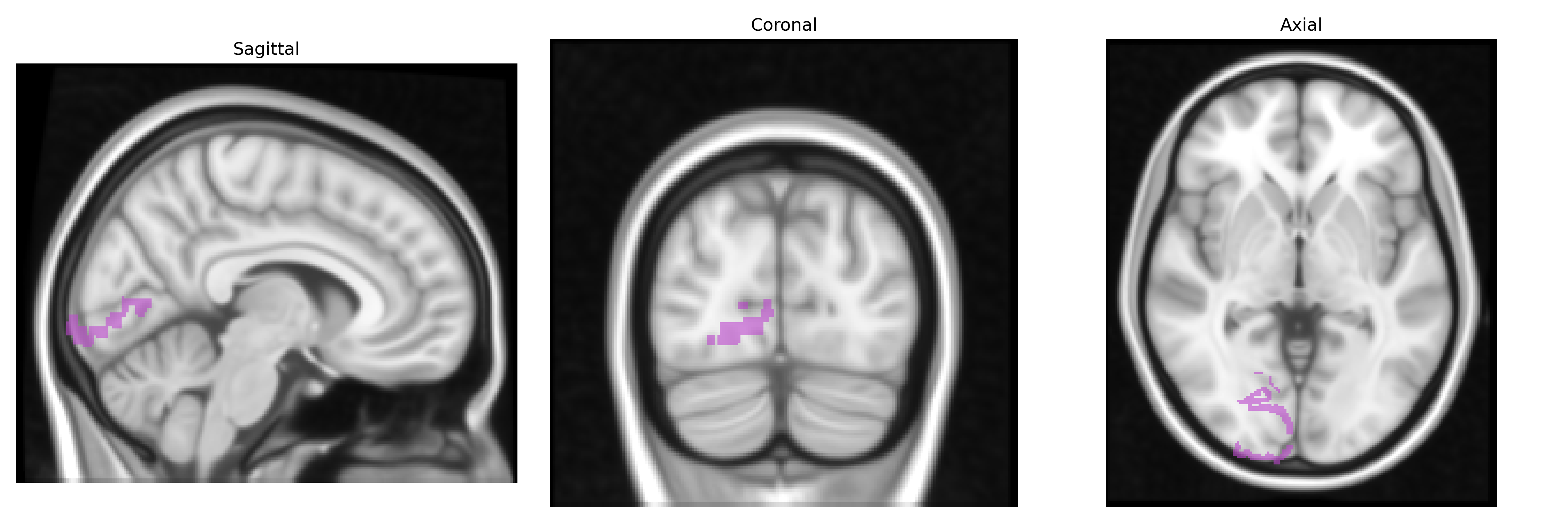
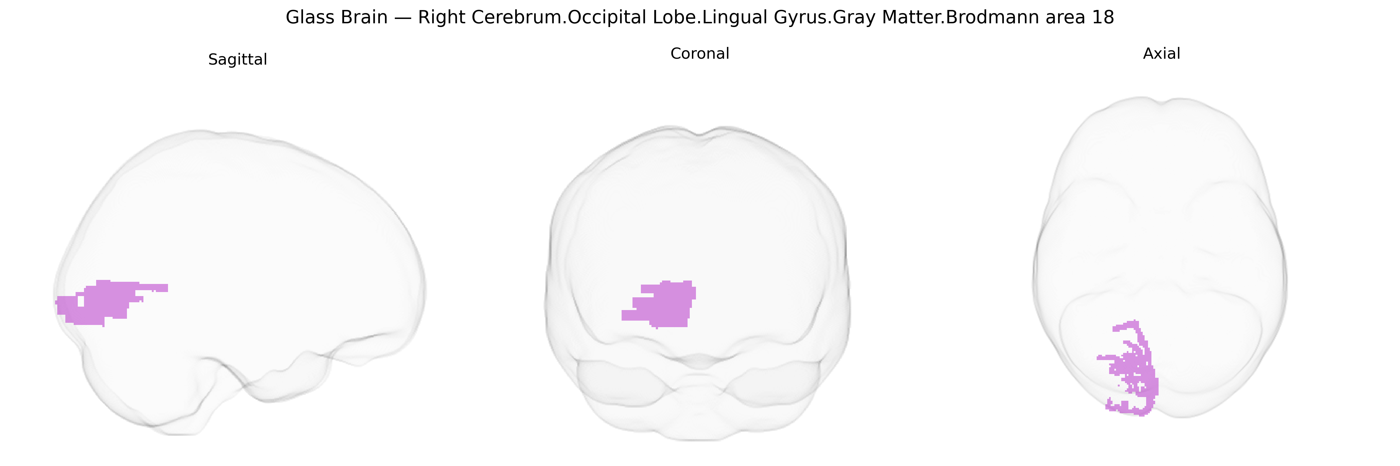
The bilateral Right Cerebrum.Occipital Lobe.Lingual Gyrus.Gray Matter.Brodmann area 18 corresponds to a portion of the secondary visual cortex (V2) located on the ventral medial surface of the occipital lobe within the lingual gyrus, immediately anterior to primary visual cortex (Brodmann area 17). This region participates in early-stage visual processing beyond primary edge and contrast detection, contributing to the integration of binocular visual inputs, orientation and spatial frequency analysis, and the formation of more complex visual representations that support object, form, and pattern perception. Neuronal populations in this area exhibit a high degree of retinotopic organization, maintaining a systematic mapping of the visual field, and receive dense afferent projections from area 17 while sending outputs to higher-order visual association areas in the occipital and temporal lobes. There is no direct link for this exact composite region; a related structure entry is Brodmann area 18.
The bilateral right lingual gyrus gray matter in Brodmann area 18, as defined in the Talairach 1 mm atlas, has been implicated in multiple genetic and GWAS findings primarily related to visual processing, neurodevelopment, and psychiatric traits: common variants in visual system and synaptic genes (including those affecting occipital cortical thickness and surface area) have shown associations with structural measures of the lingual gyrus in large imaging–genetics consortia (e.g., ENIGMA), often highlighting genes involved in neuronal migration, axon guidance, and glutamatergic signaling; polygenic risk scores for schizophrenia, bipolar disorder, major depression, and autism spectrum disorder have been linked to reduced gray matter volume or altered activation in this region, suggesting that pleiotropic psychiatric risk variants contribute to occipital–lingual structural variation; GWAS of migraine, especially visual or aura-related subtypes, report associations with occipital cortical areas that include or border BA18, while visual acuity and refractive error GWAS (e.g., for myopia) have identified genetic influences that partly map onto occipital cortical thickness, including the lingual gyrus; in addition, genetic variants associated with reading, dyslexia, and general cognitive ability show imaging correlates in secondary visual cortices such as BA18, consistent with the role of the lingual gyrus in processing letters, words, and complex visual patterns; finally, Alzheimer’s disease, Parkinson’s disease, and multiple sclerosis risk loci have been associated with downstream atrophy or microstructural alterations encompassing the lingual gyrus, though these effects are generally less specific and reflect broader neurodegenerative or demyelinating processes rather than a uniquely targeted genetic influence on this single subregion.
Overview generated by GPT-4o (2026).
Region ID: 249
Hemisphere: bilateral
Atlas: Talairach labels 1mm
Full Quality Version: Download MP4
Full Quality Version: Download MP4
Wali Sidiqyar*, Gaurav Rudravaram*, Elyssa M. McMaster, Trent M. Schwartz, Adam M. Saunders, Kurt G. Schilling, Bennett A. Landman "Introducing SPINS: A Shared Public Visualization Library of Neuroanatomical Structures." Medical Imaging with Deep Learning- short paper
This resource is licensed under CC0 1.0 Universal (Public Domain).