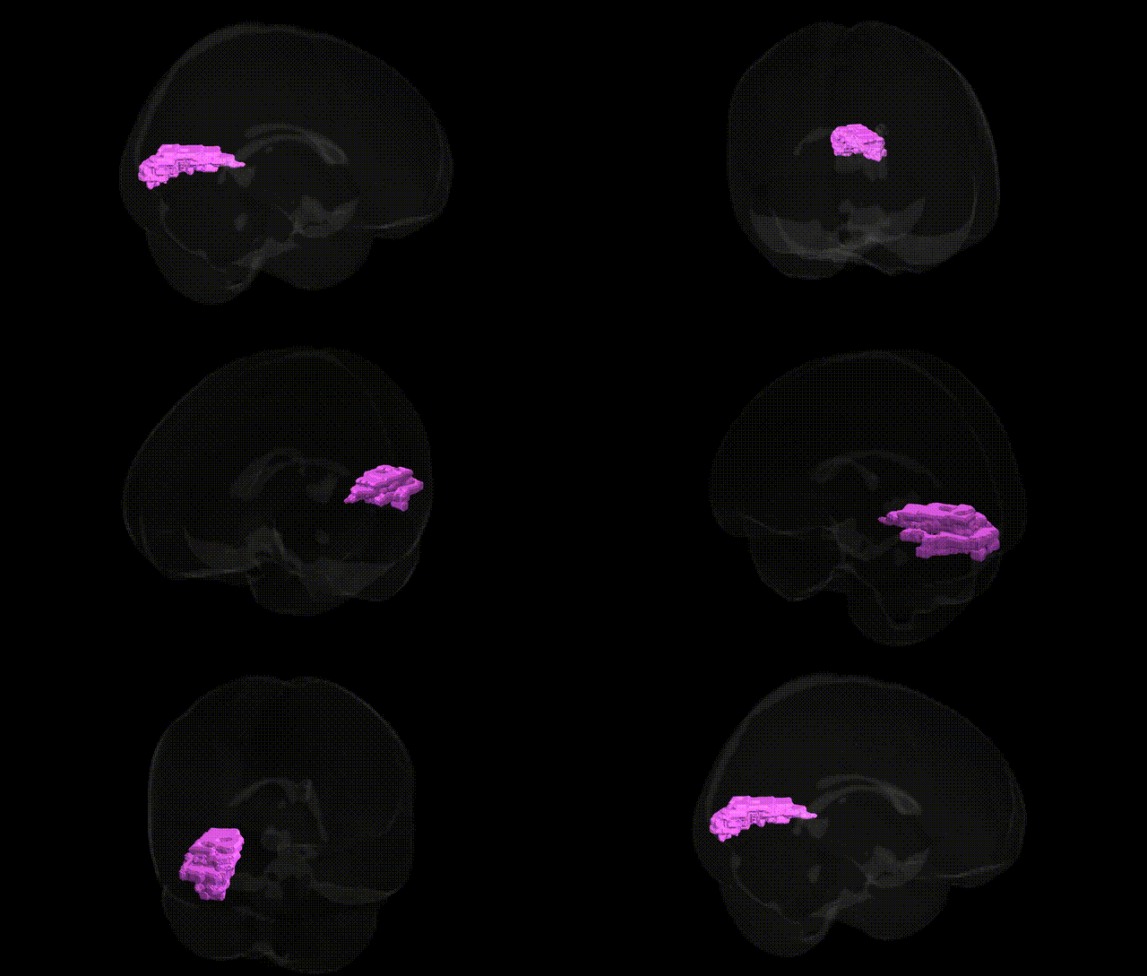
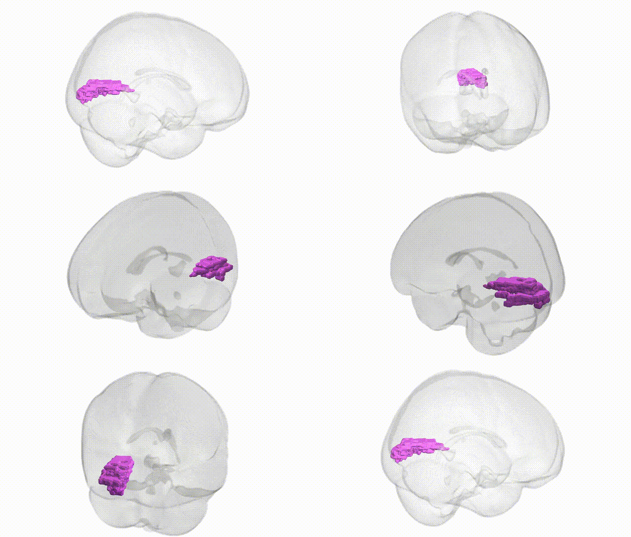
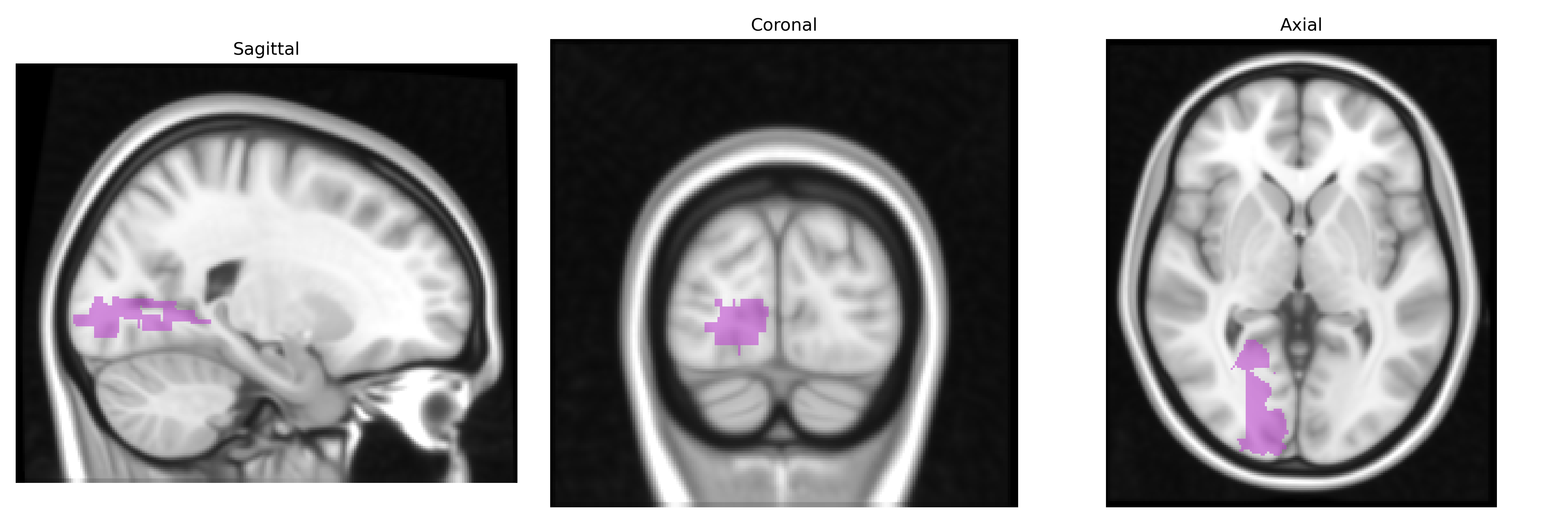
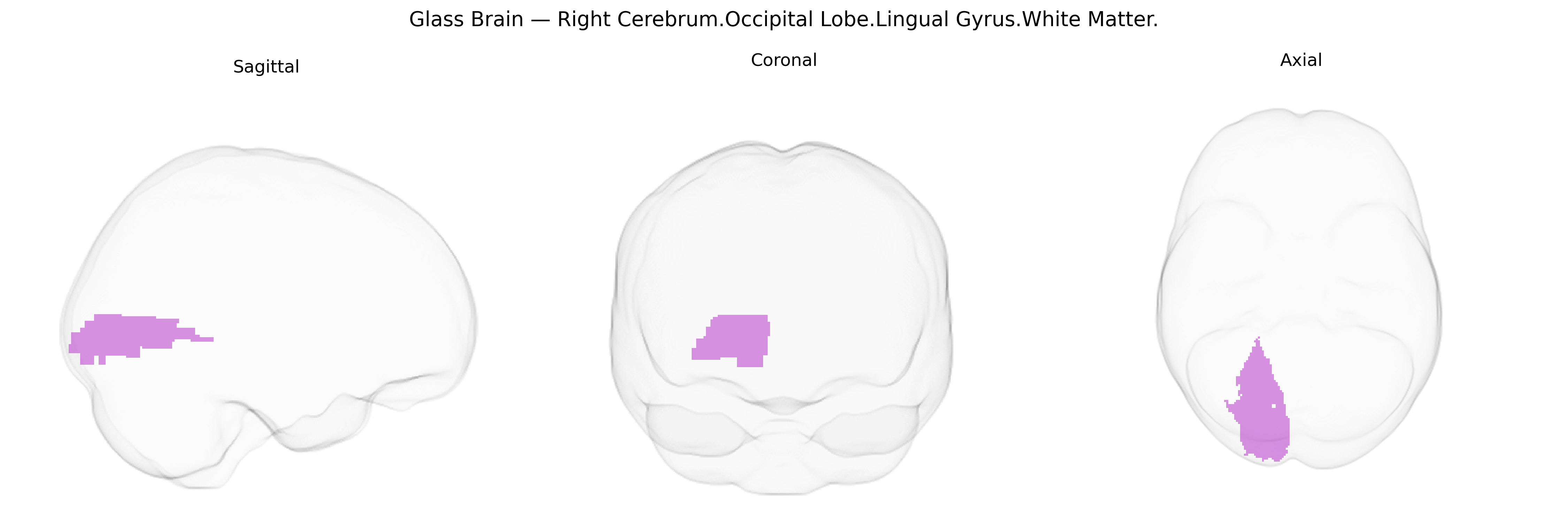
The bilateral Right Cerebrum Occipital Lobe Lingual Gyrus White Matter comprises the subcortical fiber tracts underlying the lingual gyrus in the occipital lobe, a region situated on the ventral surface of the occipital cortex and extending into the medial occipito-temporal area. This white matter contains association and projection fibers that interconnect primary and secondary visual cortices with downstream visual processing regions implicated in complex visual functions, including aspects of visual recognition, processing of letters and words, and components of visual memory. These fibers contribute to ventral visual stream pathways that support form and object perception, as well as integration of visual inputs with higher-order cognitive processes. There is no direct link for the white matter of the lingual gyrus; a related article is Lingual gyrus.
The bilateral right cerebrum occipital lingual gyrus white matter, as defined by the Talairach 1 mm atlas, lies within the occipital-temporo‑medial visual pathway and has been implicated in several GWAS and imaging-genetics studies that examine white matter microstructure, cortical connectivity, and occipital lobe volume. Variants in genes related to axonal guidance, myelination, and neurodevelopment (for example, loci near or within NTRK3, CNTNAP2, and several oligodendrocyte-related genes) have been associated with diffusion MRI measures (e.g., fractional anisotropy and mean diffusivity) in occipital and lingual gyrus white matter tracts, though most findings are not specific to this gyrus alone and arise from large-scale, atlas-based or tract-based analyses (e.g., UK Biobank–derived GWAS of white matter integrity). Genetic risk for neurodevelopmental and psychiatric disorders—including autism spectrum disorder, schizophrenia, major depression, and attention-deficit/hyperactivity disorder—has been linked to altered occipital/lingual white matter via polygenic risk score and ENIGMA-type consortia analyses, indicating that distributed risk loci affecting synaptic plasticity, glutamatergic signaling, and myelin structure contribute to microstructural changes in this region. In addition, genetic variants associated with visual processing traits and reading-related phenotypes (such as dyslexia risk loci in or near DCDC2 and KIAA0319) show convergent evidence of altered occipital and lingual connectivity, although spatial localization at 1 mm Talairach resolution is approximate. Overall, current genetic associations for the bilateral right lingual gyrus white matter derive largely from broad imaging-genetics and polygenic studies rather than region-specific GWAS, with most signals reflecting shared neurodevelopmental and connectivity-related genetic architecture across posterior visual white matter tracts.
Overview generated by GPT-4o (2026).
Region ID: 253
Hemisphere: bilateral
Atlas: Talairach labels 1mm
Full Quality Version: Download MP4
Full Quality Version: Download MP4
Wali Sidiqyar*, Gaurav Rudravaram*, Elyssa M. McMaster, Trent M. Schwartz, Adam M. Saunders, Kurt G. Schilling, Bennett A. Landman "Introducing SPINS: A Shared Public Visualization Library of Neuroanatomical Structures." Medical Imaging with Deep Learning- short paper
This resource is licensed under CC0 1.0 Universal (Public Domain).