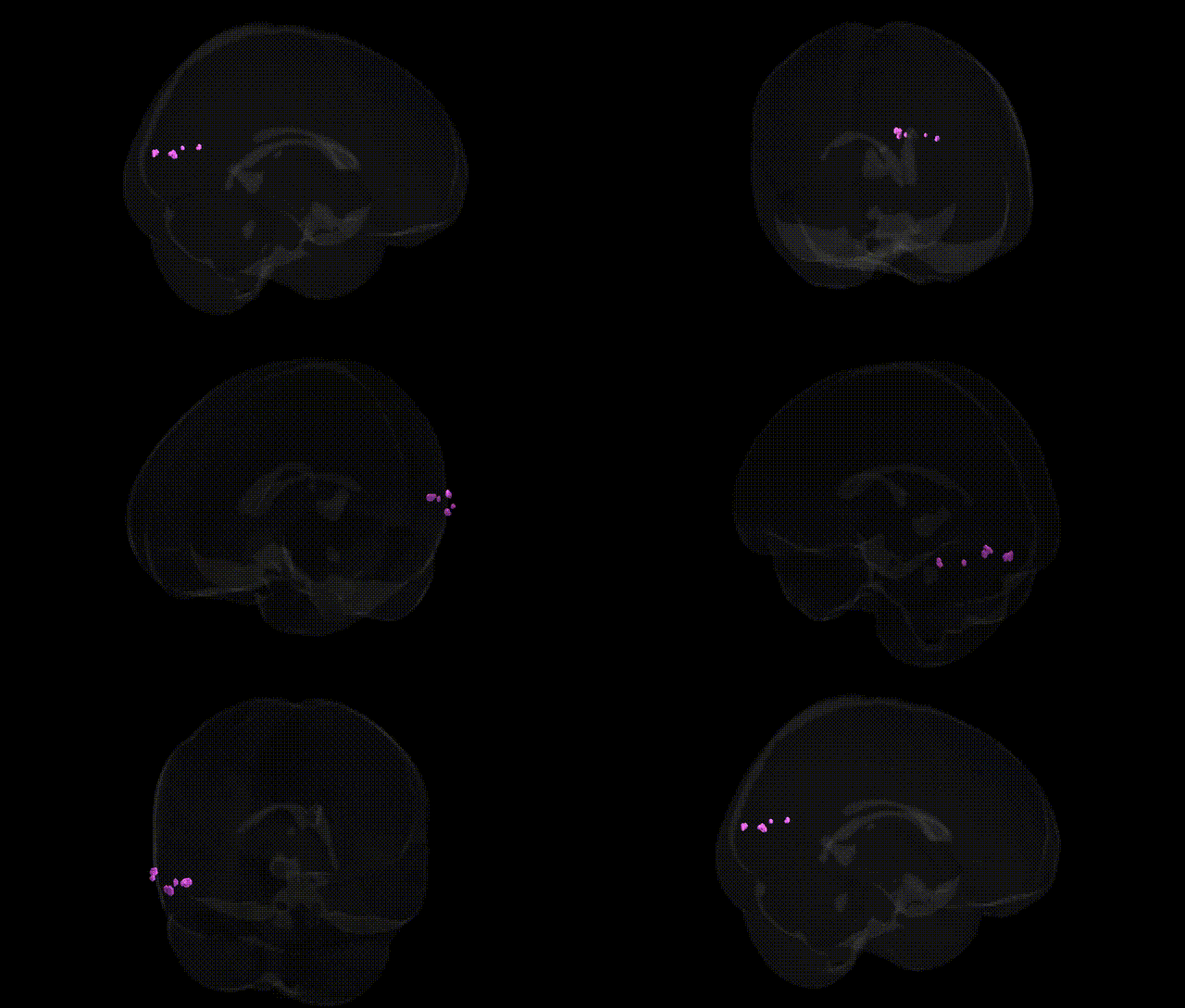
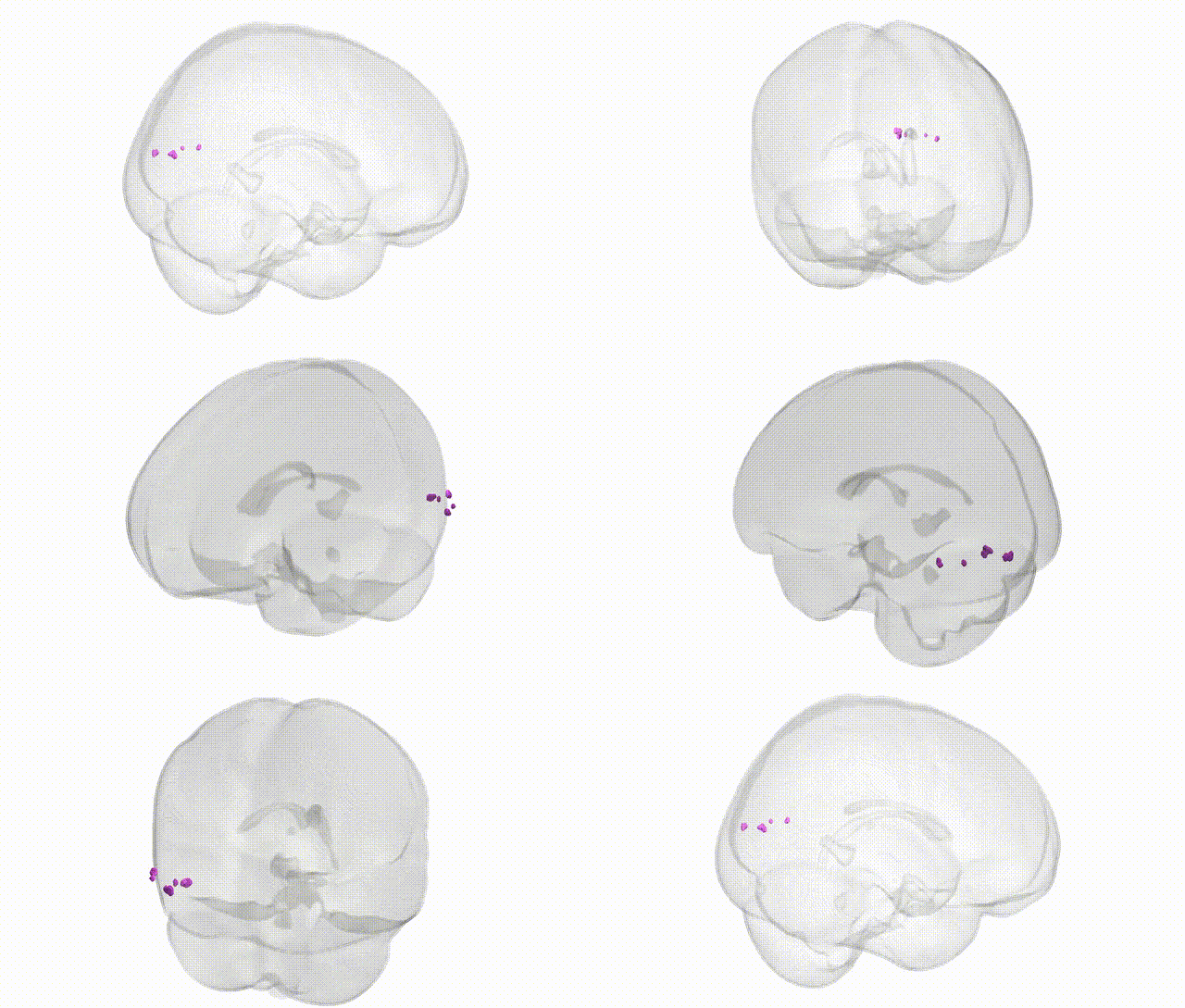
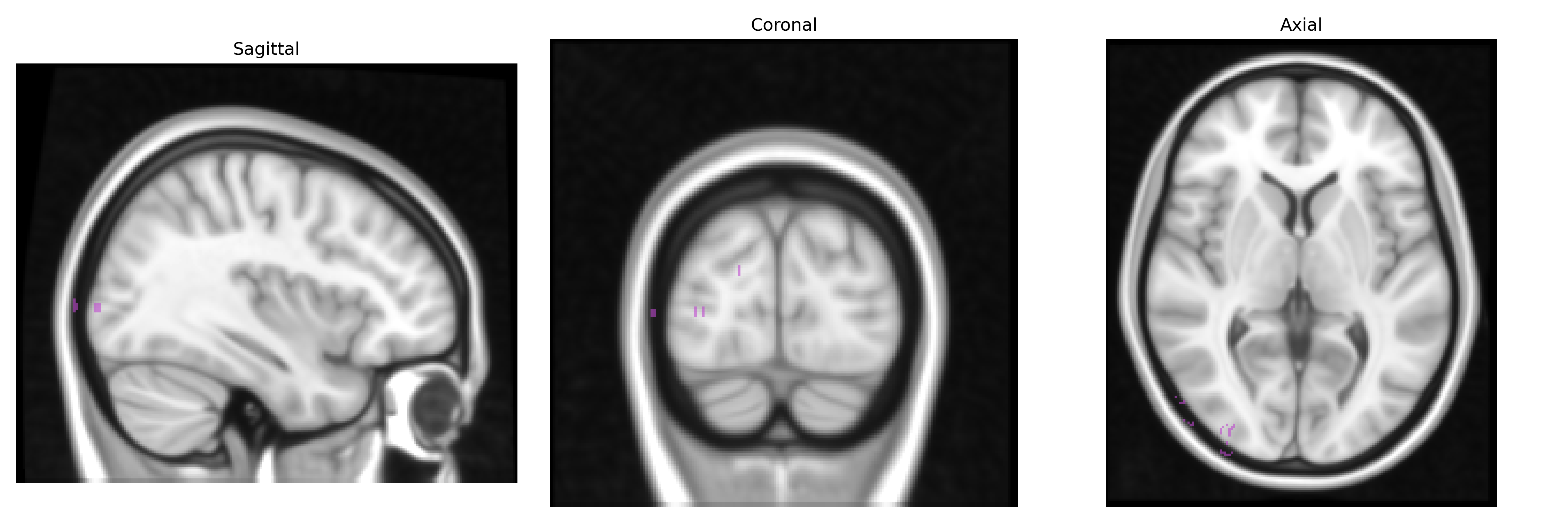
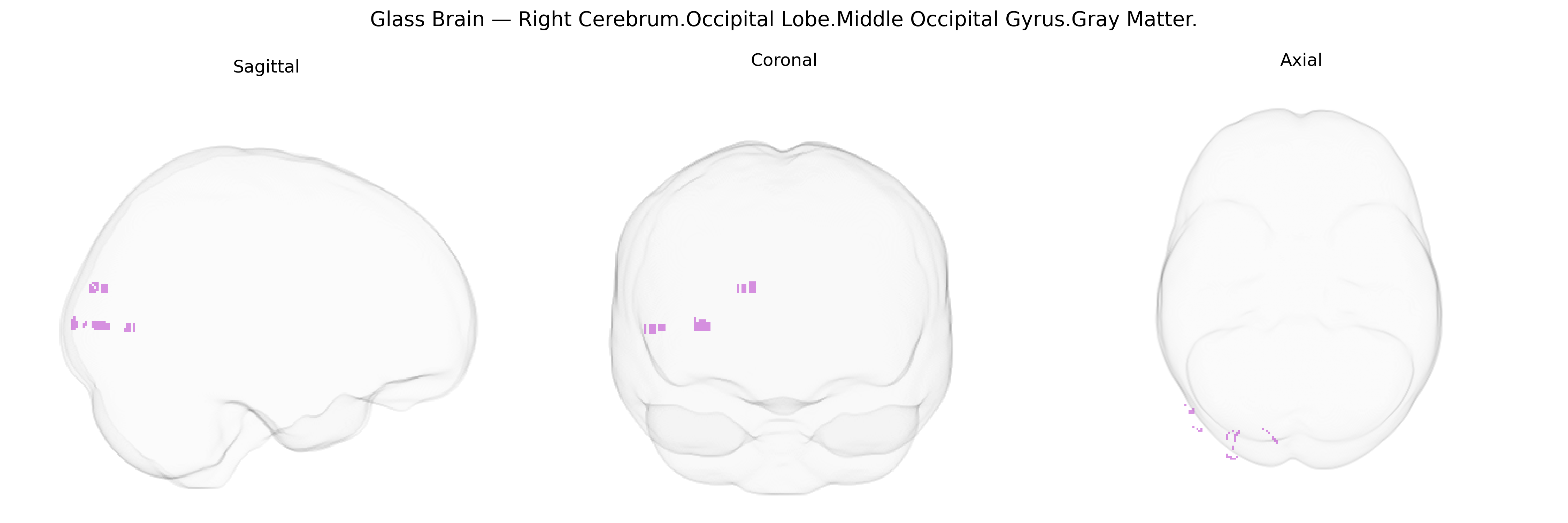
The bilateral Right Cerebrum.Occipital Lobe.Middle Occipital Gyrus.Gray Matter corresponds to cortical gray matter in the middle portion of the occipital lobe on the right hemisphere, encompassing regions involved in intermediate stages of visual processing. This gyrus lies between the superior and inferior occipital gyri and participates in analyzing motion, shape, and spatial aspects of visual stimuli, integrating information from primary visual cortex (V1) and projecting to higher-order visual association areas within the dorsal and ventral visual streams. Functionally, it contributes to object recognition, visuospatial perception, and the coordination of visual input with attention and eye movements. There is no direct link; see Occipital lobe.
The bilateral right middle occipital gyrus gray matter, as defined in the Talairach 1 mm atlas, has been implicated in several genetic and imaging-genetics studies, particularly through large-scale GWAS of cortical thickness and surface area that identify common variants in genes regulating neurodevelopment, synaptic function, and visual processing; loci near genes such as MAPT, WNT signaling components, and cell-adhesion or axon-guidance genes have shown associations with occipital lobe morphology, though effects are typically small and regionally overlapping. Occipital and specifically middle occipital gyrus measures have also emerged in polygenic risk analyses for psychiatric and neurodevelopmental disorders, including schizophrenia, major depression, autism spectrum disorder, and ADHD, where risk alleles correlate with altered gray matter volume or thickness in visual association cortex; in addition, migraine, visual hallucinations in Parkinson’s disease, and some forms of epilepsy show structural or functional abnormalities in this region that co-localize with disorder-associated genetic variants or polygenic burden. Nonetheless, the existing literature generally treats the middle occipital gyrus as part of a broader occipital/visual network rather than a genetically isolated parcel, so current genetic associations are best interpreted as reflecting distributed effects on visual cortical development and connectivity rather than specific, well-replicated, gene–region relationships confined uniquely to the right middle occipital gyrus.
Overview generated by GPT-4o (2026).
Region ID: 521
Hemisphere: bilateral
Atlas: Talairach labels 1mm
Full Quality Version: Download MP4
Full Quality Version: Download MP4
Wali Sidiqyar*, Gaurav Rudravaram*, Elyssa M. McMaster, Trent M. Schwartz, Adam M. Saunders, Kurt G. Schilling, Bennett A. Landman "Introducing SPINS: A Shared Public Visualization Library of Neuroanatomical Structures." Medical Imaging with Deep Learning- short paper
This resource is licensed under CC0 1.0 Universal (Public Domain).