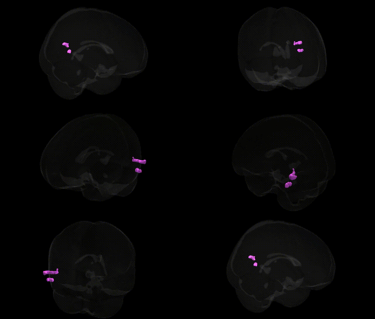
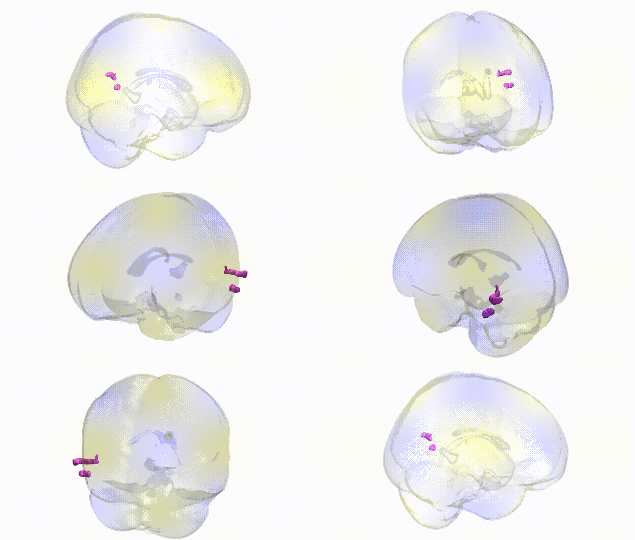
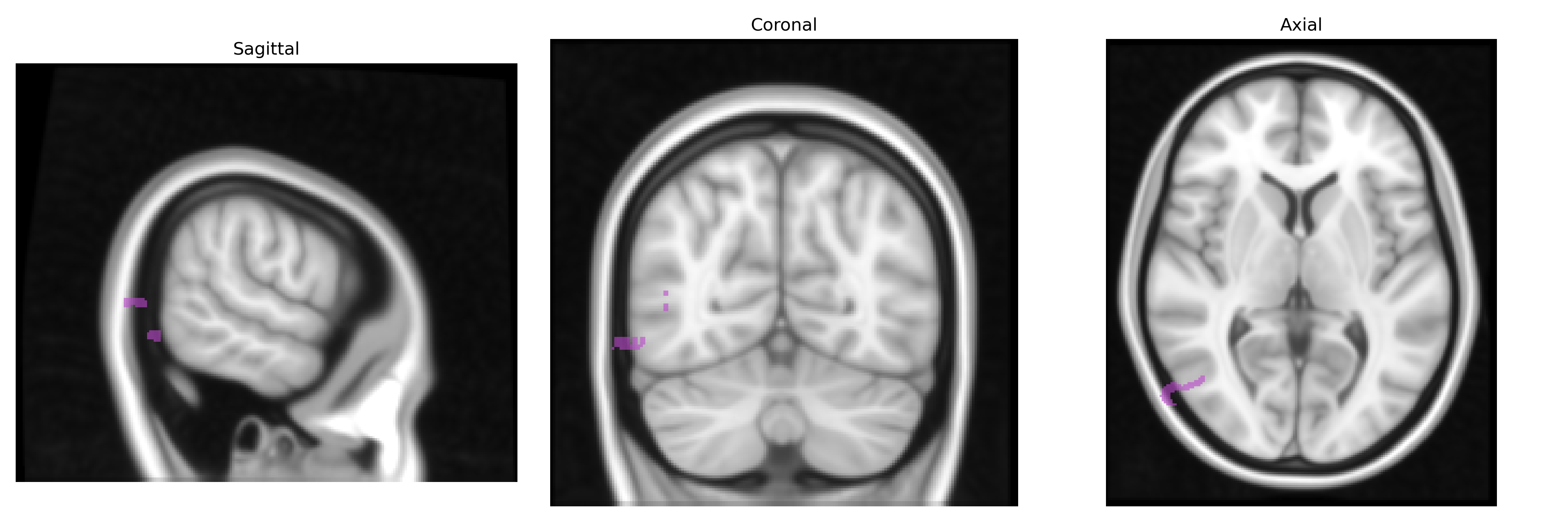
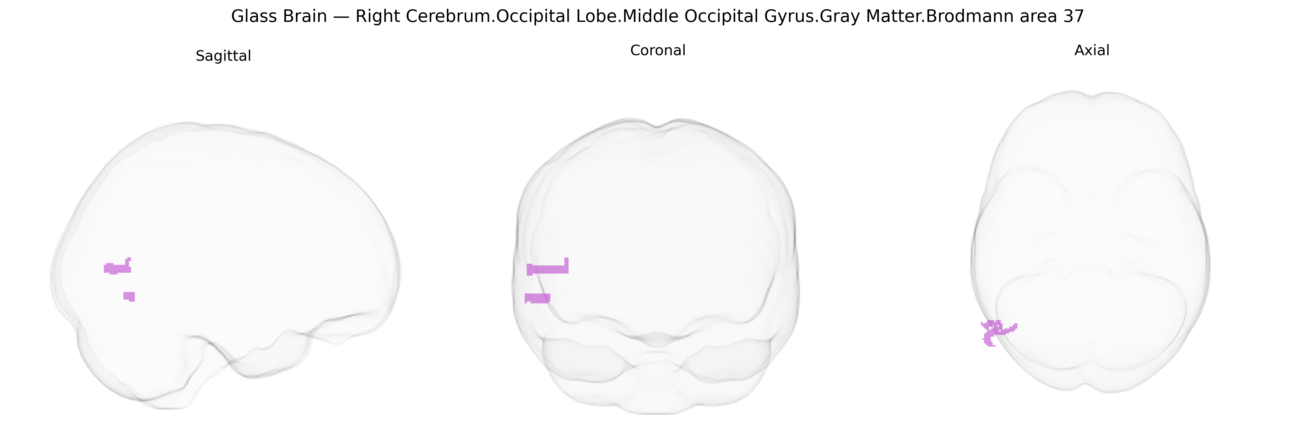
The bilateral Right Cerebrum.Occipital Lobe.Middle Occipital Gyrus.Gray Matter.Brodmann area 37 corresponds to a ventral occipitotemporal association region implicated in high-level visual processing, particularly object, face, and word-form recognition, as well as aspects of semantic processing. Situated at the junction of the occipital and temporal lobes, BA37 integrates inputs from earlier visual areas (e.g., BA17–19) and conveys processed information to temporal and frontal regions involved in language and memory. Its gray matter contains columnar and laminar cortical architecture typical of association cortex, supporting complex pattern analysis and multimodal integration. Functionally, this territory overlaps with the fusiform and adjacent occipitotemporal cortex and forms part of the ventral visual (“what”) pathway. There is no direct Wikipedia article for “Brodmann area 37,” but a closely related and encompassing structure is the Fusiform gyrus.
The right middle occipital gyrus in Brodmann area 37 (part of the ventral occipitotemporal cortex/fusiform region) is strongly implicated in visual object and face processing, and genetic associations with this region largely emerge from imaging–genetics and GWAS of brain structure and function rather than region-specific single-gene studies. Variants in genes involved in neurodevelopment and synaptic plasticity, including BDNF (e.g., Val66Met), NRG1, and COMT, have been associated with occipitotemporal cortical thickness or activation patterns, while large-scale GWAS of cortical morphology (e.g., ENIGMA and UK Biobank–based studies) have identified polygenic influences on occipital and temporal lobe surface area and thickness, implicating loci near genes such as HMGA2, KIAA0586, WNT signaling components, and multiple neurodevelopmental transcription factors, though these are usually reported at lobar or regional scales rather than specifically at BA37. Functional MRI–GWAS (imaging GWAS) has linked common variants to activation in occipitotemporal regions during face and word recognition tasks, with polygenic scores for educational attainment, intelligence, and cognitive performance showing associations with structural and functional measures in this territory. Clinically, mutations and risk variants in genes associated with autism spectrum disorder (e.g., SHANK3, CNTNAP2), dyslexia (e.g., DCDC2, KIAA0319), and schizophrenia (various risk loci in MHC and synaptic genes) have been related to altered structure or activity in occipitotemporal cortex, including middle occipital/fusiform areas, supporting a polygenic and pleiotropic architecture whereby shared genetic risk influences BA37-related circuits. Overall, current evidence indicates that bilateral BA37, including the right middle occipital gyrus, is shaped by highly polygenic common variation affecting brain size, cortical patterning, and visual-cognitive networks, with disorder associations arising from broad neurodevelopmental and synaptic pathways rather than a small set of region-specific genes.
Overview generated by GPT-4o (2026).
Region ID: 324
Hemisphere: bilateral
Atlas: Talairach labels 1mm
Full Quality Version: Download MP4
Full Quality Version: Download MP4
Wali Sidiqyar*, Gaurav Rudravaram*, Elyssa M. McMaster, Trent M. Schwartz, Adam M. Saunders, Kurt G. Schilling, Bennett A. Landman "Introducing SPINS: A Shared Public Visualization Library of Neuroanatomical Structures." Medical Imaging with Deep Learning- short paper
This resource is licensed under CC0 1.0 Universal (Public Domain).