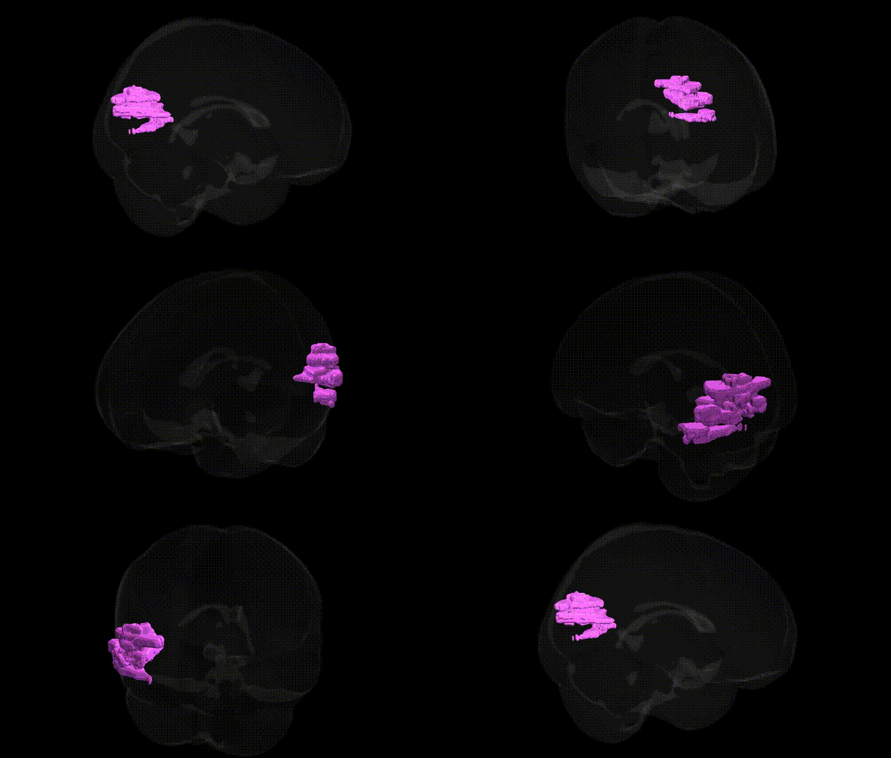
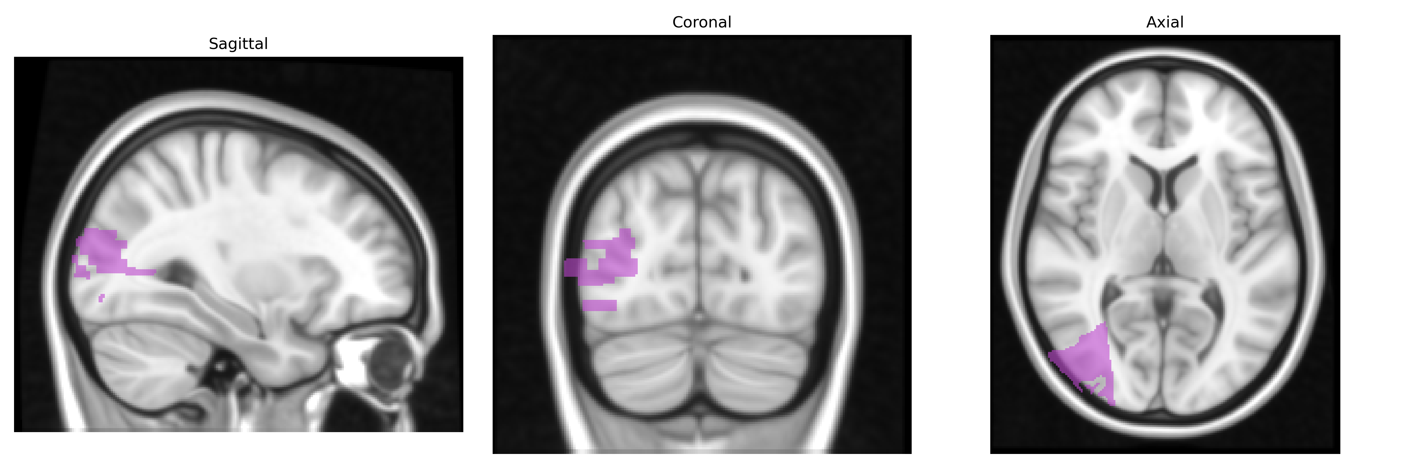
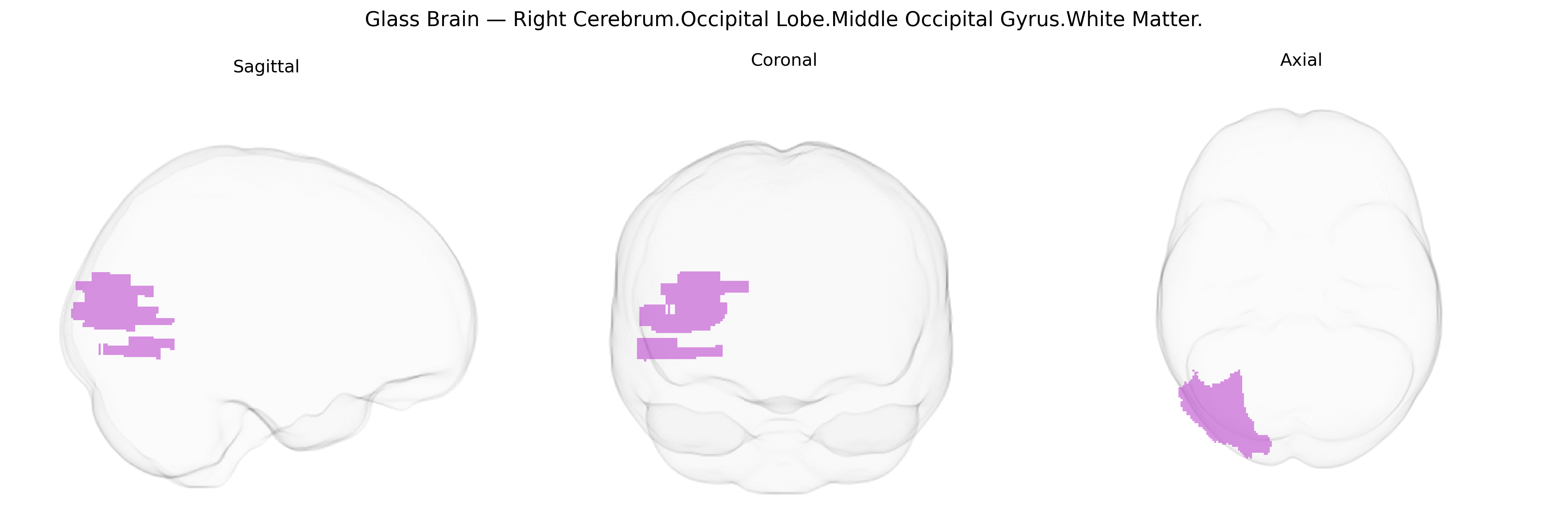
The bilateral Right Cerebrum Occipital Lobe Middle Occipital Gyrus White Matter corresponds to subcortical myelinated fiber pathways deep to the middle occipital gyrus in the lateral portion of the occipital lobe, involved in transmitting visual information between primary and higher-order visual cortices and with temporal and parietal association areas. This white matter includes components of long association tracts such as portions of the inferior longitudinal fasciculus and possibly the inferior fronto-occipital fasciculus, which support object recognition, visual scene processing, and integration of visual input with memory and spatial functions. Pathology in this region can contribute to higher-order visual deficits, including impairments in form and motion perception. There is no direct Wikipedia article for this exact white-matter label; a closely related structure is the Middle Occipital Gyrus.
Genetic associations specifically annotated to the bilateral Right Cerebrum.Occipital Lobe.Middle Occipital Gyrus.White Matter in the Talairach 1 mm atlas are not well resolved at the fine-grained regional level, but large-scale imaging genetics and GWAS of white-matter microstructure and occipital lobe morphology implicate multiple loci and pathways influencing this territory. Diffusion tensor imaging GWAS (e.g., ENIGMA, UK Biobank) have identified variants near genes involved in axon guidance, myelination, and oligodendrocyte function (such as NRG1, MAG, and other myelin-related loci) that affect fractional anisotropy and mean diffusivity in posterior association tracts traversing the middle occipital white matter, including portions of the inferior and superior longitudinal fasciculi and optic radiations. Occipital white-matter measures also share polygenic overlap with neurodevelopmental and psychiatric conditions—autism spectrum disorder, schizophrenia, major depression, and attention-deficit/hyperactivity disorder—where risk loci in synaptic and neurodevelopmental genes (e.g., CACNA1C, GRIN2A, CNTNAP2) show pleiotropic effects on both disease risk and occipital or visual-association white-matter integrity. Additional GWAS link occipital-lobe and global white-matter volume, as well as visual processing–related traits, to general neurodevelopmental loci (e.g., MAPT region, WNT and axon guidance pathway genes), suggesting that genetic influences on neuronal migration, axonal projection, and myelin maintenance contribute to inter-individual variation in the structure and connectivity of the middle occipital gyrus white matter, although no single locus has emerged as uniquely specific to this precise Talairach-defined region.
Overview generated by GPT-4o (2026).
Region ID: 309
Hemisphere: bilateral
Atlas: Talairach labels 1mm
Full Quality Version: Download MP4
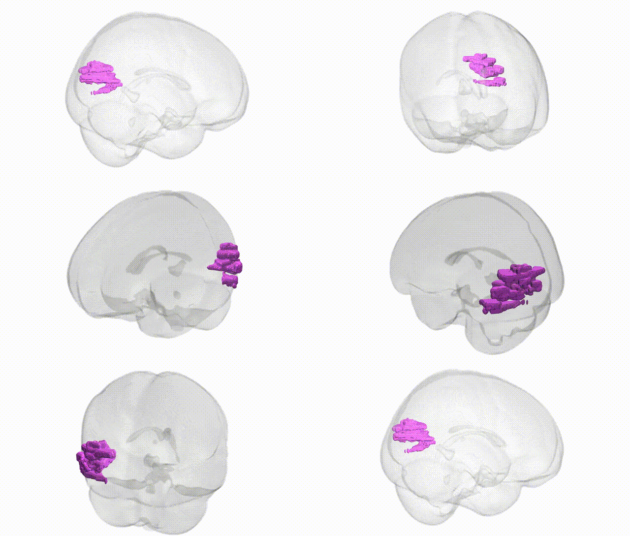
Full Quality Version: Download MP4
Wali Sidiqyar*, Gaurav Rudravaram*, Elyssa M. McMaster, Trent M. Schwartz, Adam M. Saunders, Kurt G. Schilling, Bennett A. Landman "Introducing SPINS: A Shared Public Visualization Library of Neuroanatomical Structures." Medical Imaging with Deep Learning- short paper
This resource is licensed under CC0 1.0 Universal (Public Domain).