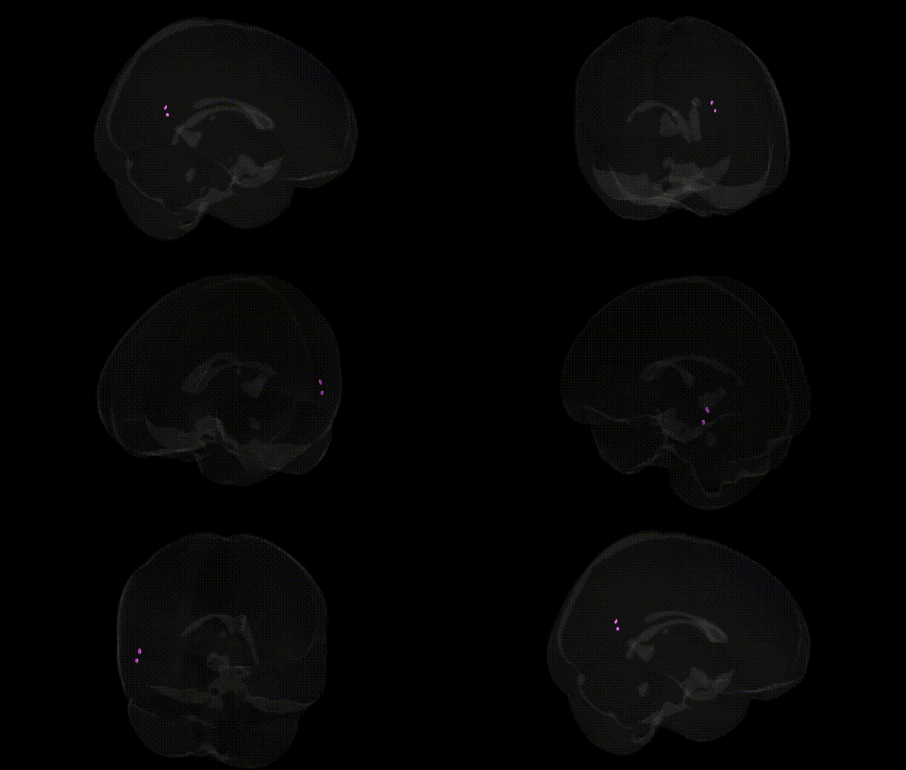
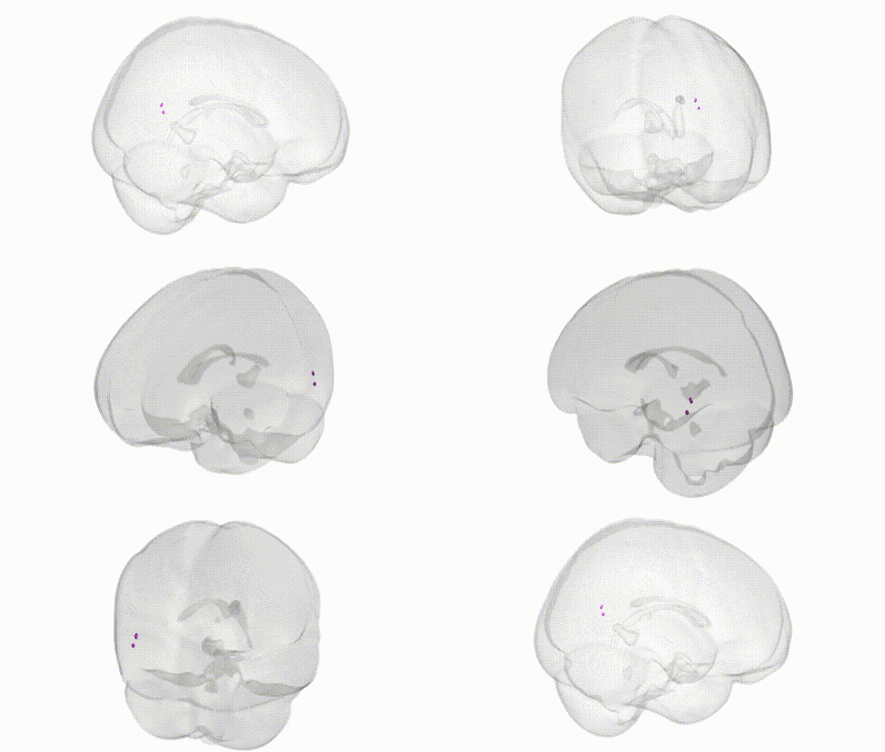
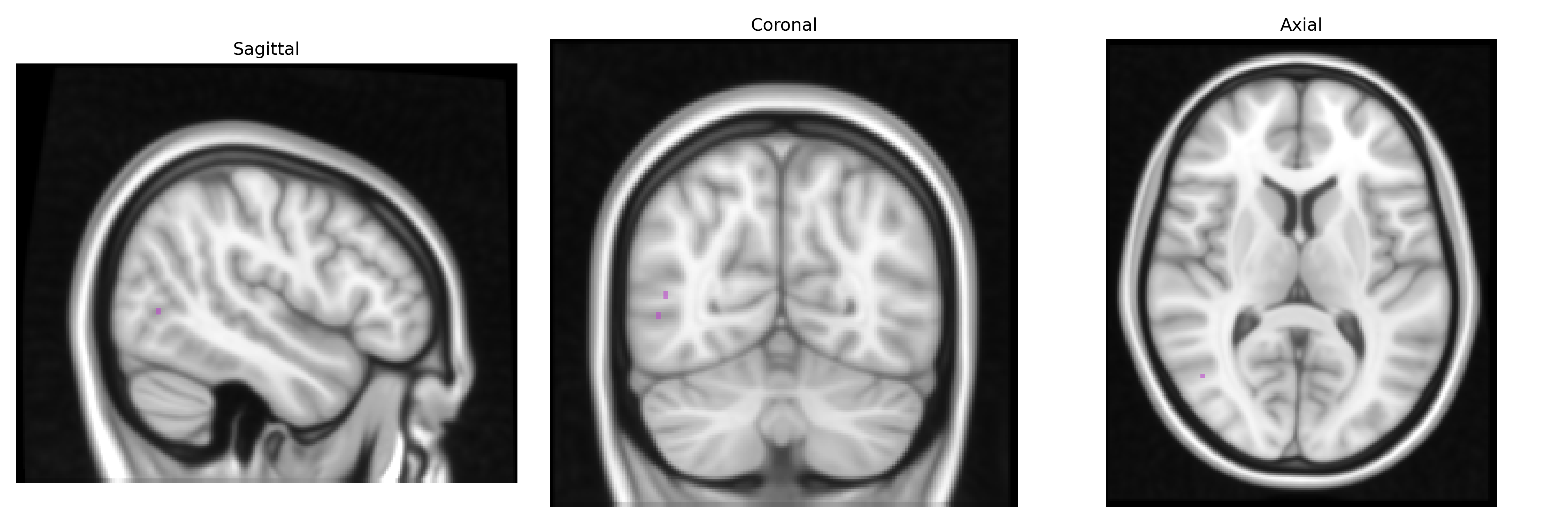
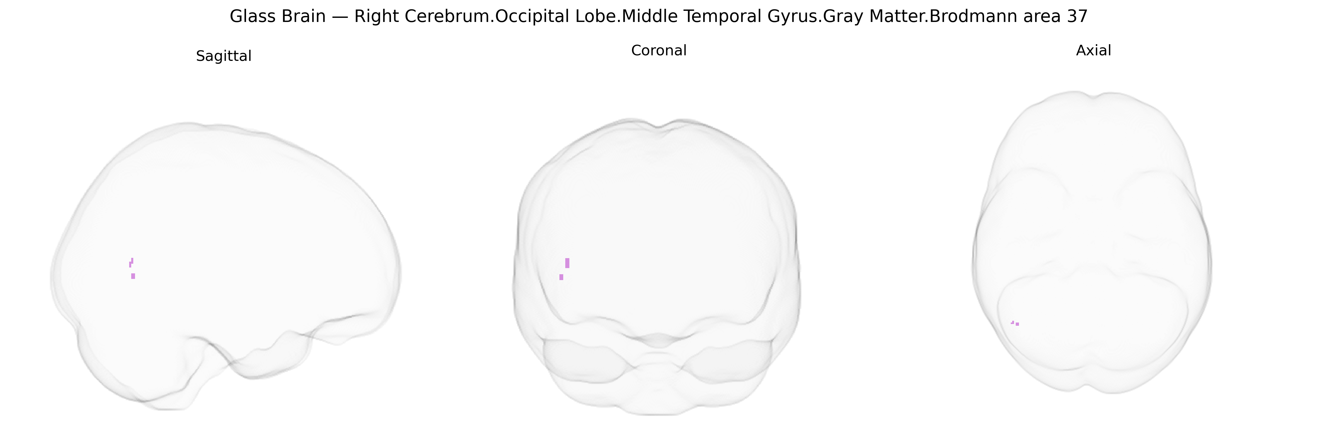
The bilateral Right Cerebrum.Occipital Lobe.Middle Temporal Gyrus.Gray Matter.Brodmann area 37 corresponds to a higher-order visual association and multimodal integration zone located at the occipitotemporal junction, overlapping the posterior and middle portions of the middle temporal gyrus and extending into the fusiform and adjacent inferior temporal cortices. This region participates in complex object, face, and word-form processing, integrating visual features with stored semantic representations and contributing to category-specific recognition (such as tools, animals, and faces), as well as aspects of visual memory and language-related functions, including naming and lexical retrieval. Neurally, Brodmann area 37 is characterized by a well-developed granular layer IV and dense corticocortical connectivity with other temporal, occipital, and inferior parietal association areas, and is frequently implicated in functional imaging and lesion studies of visual agnosia, prosopagnosia, and certain forms of aphasia. Brodmann area 37
The bilateral right occipital–temporal region corresponding to the middle temporal gyrus gray matter in Brodmann area 37 (often overlapping the fusiform and adjacent occipitotemporal cortex) has been implicated in multiple genetic and GWAS-based findings, primarily through imaging-genetics and neuropsychiatric studies rather than region-specific candidate-gene work. Large-scale imaging GWAS from consortia such as ENIGMA and UK Biobank have identified common variants near and within genes including HMGA2, IGF2BP1, and multiple loci on chromosomes 3, 6, and 17 that influence occipitotemporal cortical thickness and surface area, with BA37-related measures contributing to global patterns of genetic correlation with general cognitive ability, educational attainment, and processing speed. This region’s role in high-level visual and language-related processing (e.g., word-form and face recognition) aligns with genetic associations linking occipitotemporal morphology or activation to dyslexia and other reading disorders, where variants in genes such as DCDC2, KIAA0319, and ROBO1 show convergent evidence via altered activation or structure in left–right posterior temporal and occipitotemporal cortices, including BA37 territory. Neurodevelopmental and psychiatric GWAS (autism spectrum disorder, schizophrenia, bipolar disorder, major depression) have reported polygenic effects on temporal and occipital cortical volumes, with schizophrenia- and ASD-related polygenic risk scores correlating with reduced gray matter or altered functional connectivity in right occipitotemporal regions encompassing BA37. Face processing and fusiform-related traits show heritable variance, and twin and family studies indicate substantial heritability (often 40–70%) for occipitotemporal cortical thickness and volume, implying a significant polygenic architecture even where specific loci remain incompletely mapped. Overall, genetic associations for BA37 emphasize polygenic influences on cortical morphology and connectivity, with links to reading and language traits, visual recognition functions, and vulnerability to neurodevelopmental and psychiatric disorders, though precise, region-exclusive risk variants are not yet firmly established.
Overview generated by GPT-4o (2026).
Region ID: 560
Hemisphere: bilateral
Atlas: Talairach labels 1mm
Full Quality Version: Download MP4
Full Quality Version: Download MP4
Wali Sidiqyar*, Gaurav Rudravaram*, Elyssa M. McMaster, Trent M. Schwartz, Adam M. Saunders, Kurt G. Schilling, Bennett A. Landman "Introducing SPINS: A Shared Public Visualization Library of Neuroanatomical Structures." Medical Imaging with Deep Learning- short paper
This resource is licensed under CC0 1.0 Universal (Public Domain).