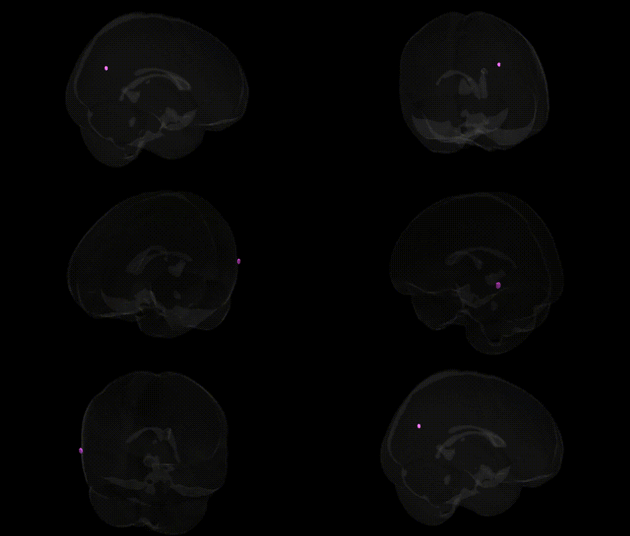
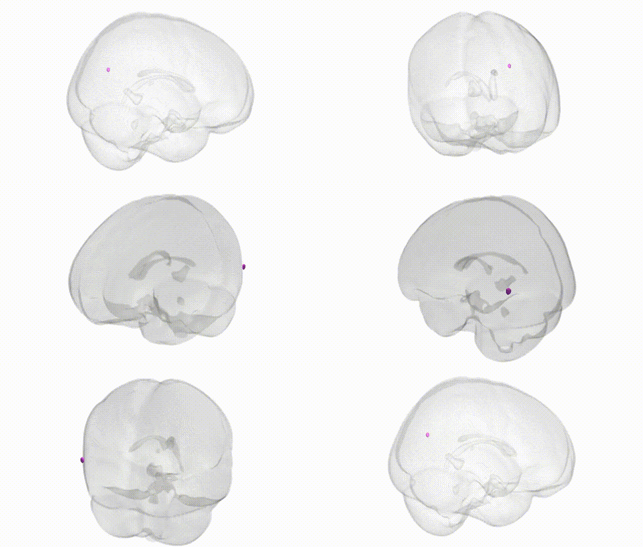
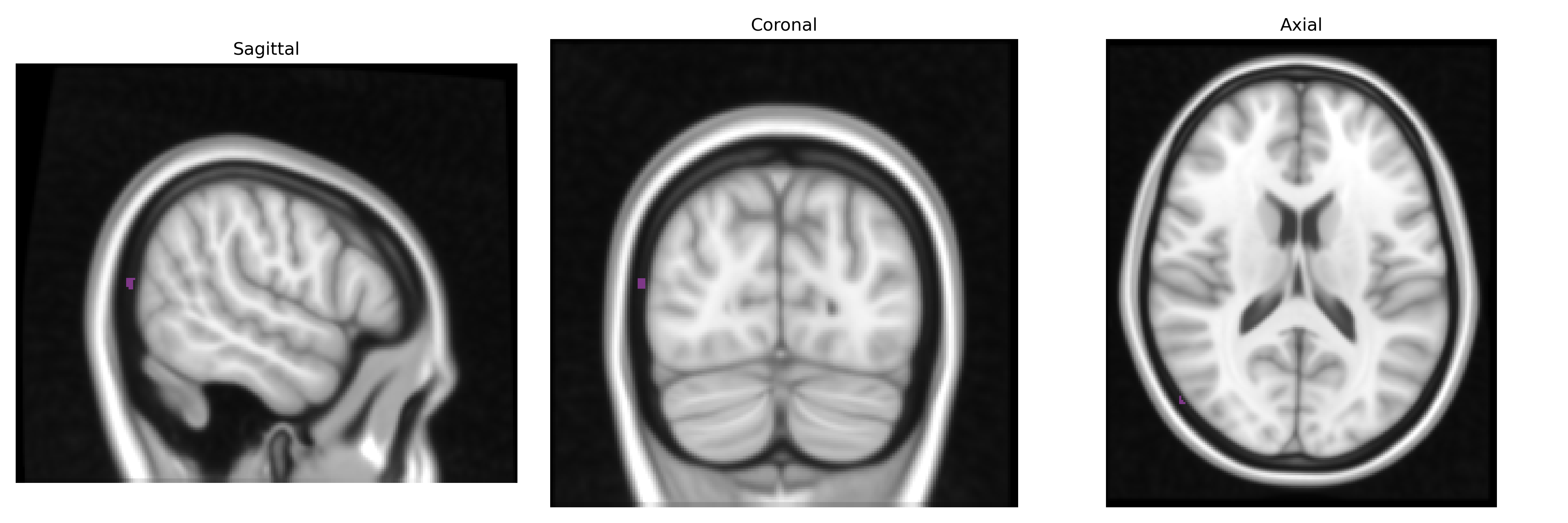
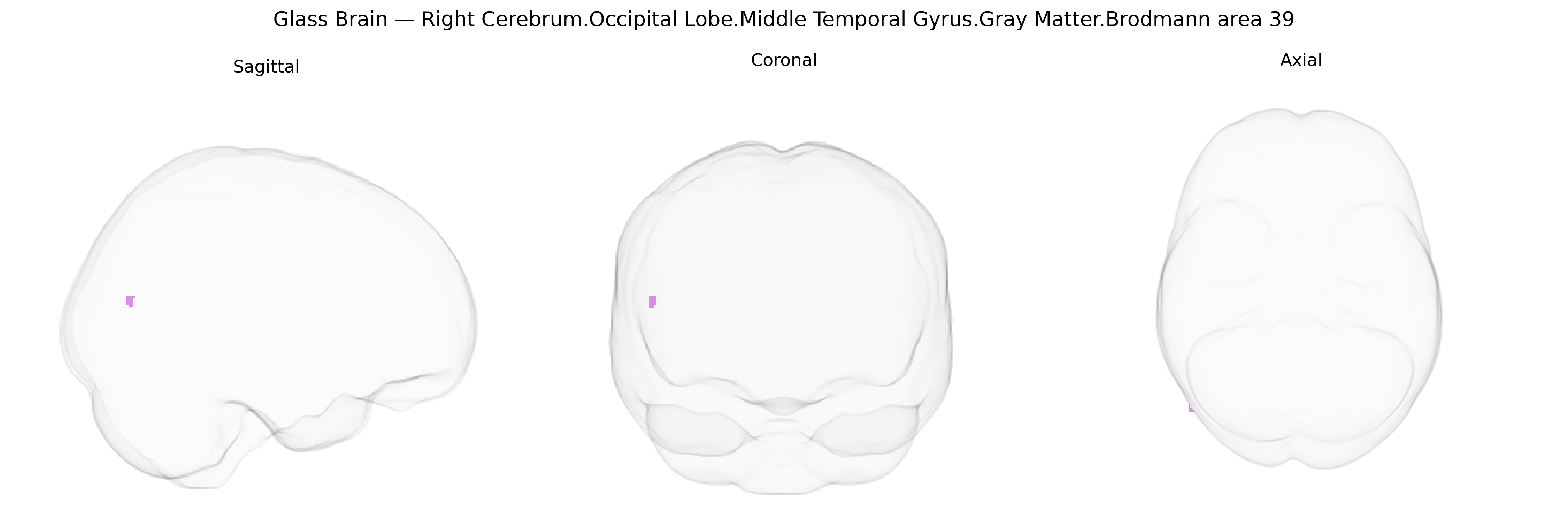
The bilateral Right Cerebrum.Occipital Lobe.Middle Temporal Gyrus.Gray Matter.Brodmann area 39 corresponds to the right-hemispheric portion of the angular gyrus region, a multimodal association cortex located at the junction of the occipital, parietal, and temporal lobes. In the Talairach 1 mm Atlas, this area lies within the gray matter of the middle temporal gyrus extending posteriorly into the occipital territory, and is implicated in high-level integrative functions including language-related processing, semantic integration, reading and writing, spatial cognition, and aspects of number processing. Neuronal populations in BA39 receive convergent visual, auditory, and somatosensory inputs, supporting the transformation of perceptual information into conceptual and symbolic representations. Functional imaging and lesion studies associate this region with components of the default mode network and with cognitive operations such as theory of mind, memory retrieval, and certain aspects of attention and visuospatial integration. There is no direct link for this exact composite Talairach label; a closely related structure is the Angular gyrus.
The bilateral right occipito-temporal region corresponding to gray matter in Brodmann area 39 (angular gyrus/posterior middle temporal–inferior parietal junction) has been repeatedly implicated in genetic studies of higher cognition and neuropsychiatric risk, although most findings derive from imaging–genetics or GWAS of cortical structure rather than this Talairach-defined parcel specifically. Large-scale cortical thickness and surface area GWAS (e.g., ENIGMA, UK Biobank–based studies) have identified common variants in genes involved in neurodevelopment and synaptic function—such as those near HMGA2, MIR2113, WNT and FGF signaling loci, and multiple 3p, 6q, and 17q regions—associated with interindividual differences in parietal/temporal association cortex, including BA39. Polygenic risk scores for educational attainment and general cognitive ability show positive associations with thicker or more voluminous inferior parietal and angular gyrus regions, while schizophrenia, major depressive disorder, and autism spectrum disorder polygenic risk scores are linked to structural or functional alterations in this area. BA39 has also emerged in language and reading-related genetic work: variants in or near genes such as KIAA0319, DCDC2, and CNTNAP2, initially identified in dyslexia and language disorders, show imaging–genetic correlations with posterior temporal–parietal regions involved in reading and semantic processing that overlap BA39, and FOXP2-regulated networks have been functionally connected with this territory. Additionally, Alzheimer’s disease risk loci (e.g., APOE, CLU, PICALM and other GWAS hits) influence atrophy patterns and amyloid-related changes that prominently involve posterior association cortex, including right angular gyrus, and small imaging–genetic studies have linked APOE ε4 and other AD polygenic risk markers to reduced gray matter and metabolic alterations in this area. Collectively, genetic associations for this region converge on pathways related to cortical development, synaptic plasticity, language and semantic networks, and vulnerability to psychiatric and neurodegenerative disorders, although resolution at the level of the precise Talairach 1 mm “Right Cerebrum.Occipital Lobe.Middle Temporal Gyrus.Gray Matter.Brodmann area 39” label remains indirect and typically inferred from broader parietal–temporal association cortex analyses.
Overview generated by GPT-4o (2026).
Region ID: 705
Hemisphere: bilateral
Atlas: Talairach labels 1mm
Full Quality Version: Download MP4
Full Quality Version: Download MP4
Wali Sidiqyar*, Gaurav Rudravaram*, Elyssa M. McMaster, Trent M. Schwartz, Adam M. Saunders, Kurt G. Schilling, Bennett A. Landman "Introducing SPINS: A Shared Public Visualization Library of Neuroanatomical Structures." Medical Imaging with Deep Learning- short paper
This resource is licensed under CC0 1.0 Universal (Public Domain).