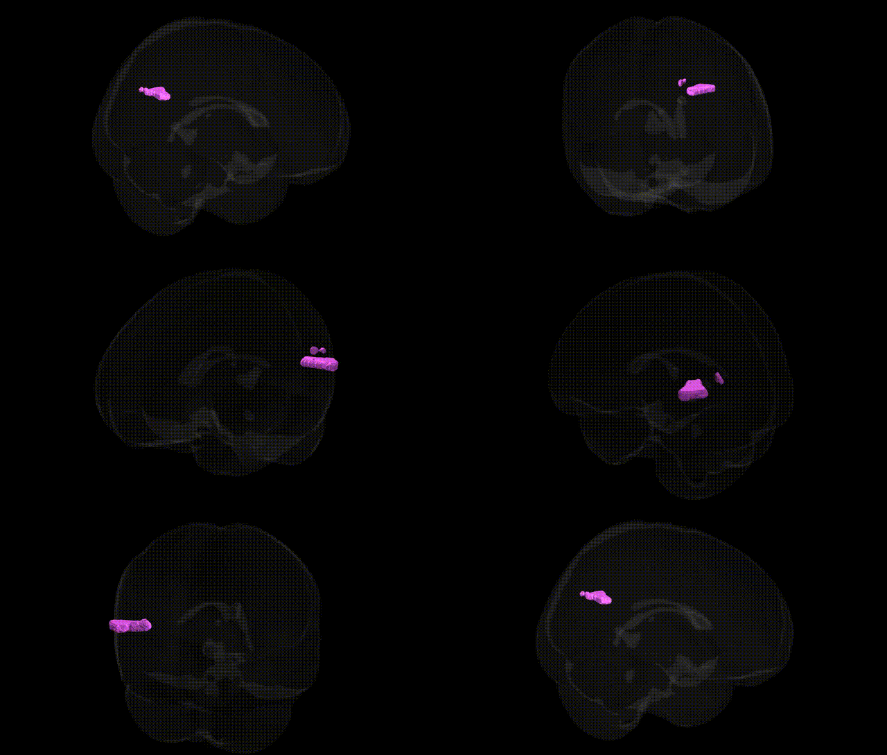
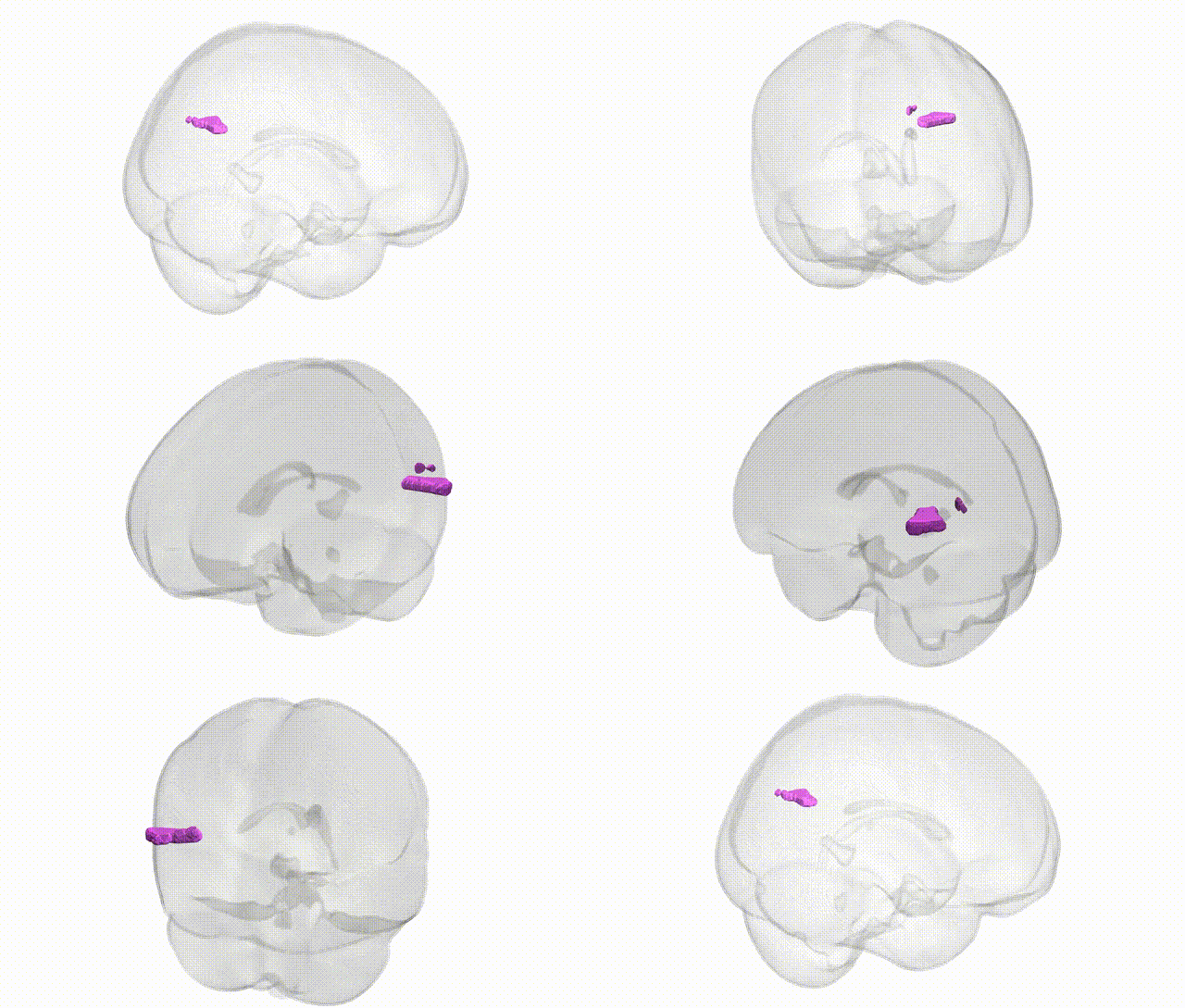
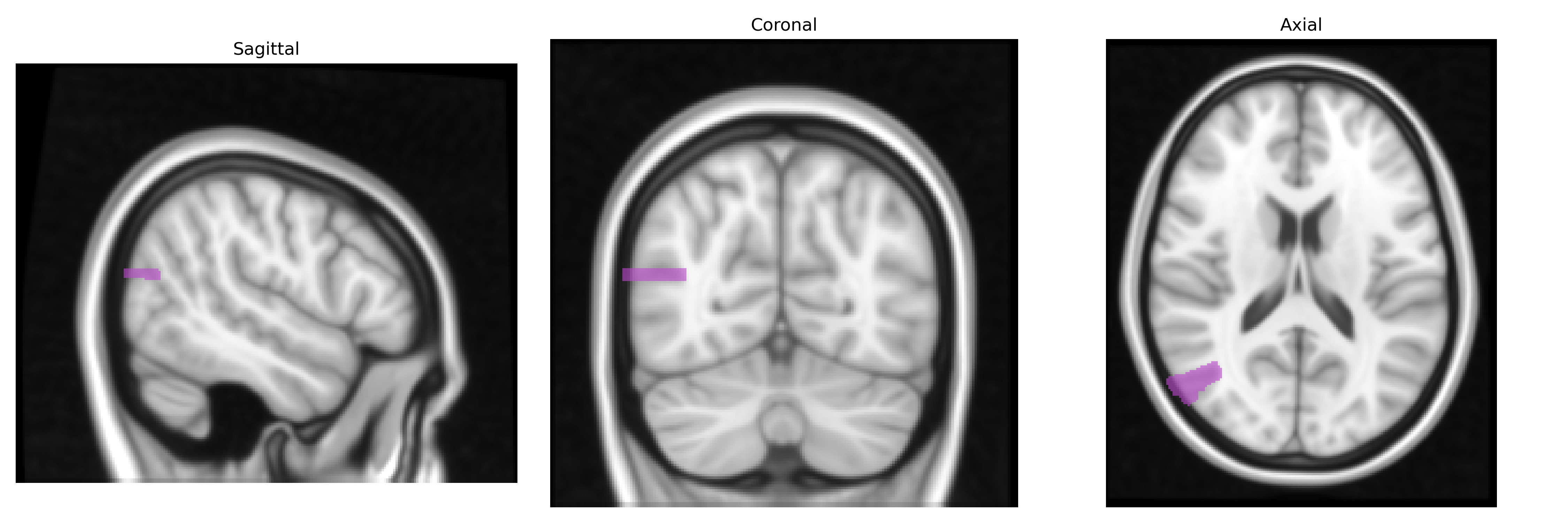
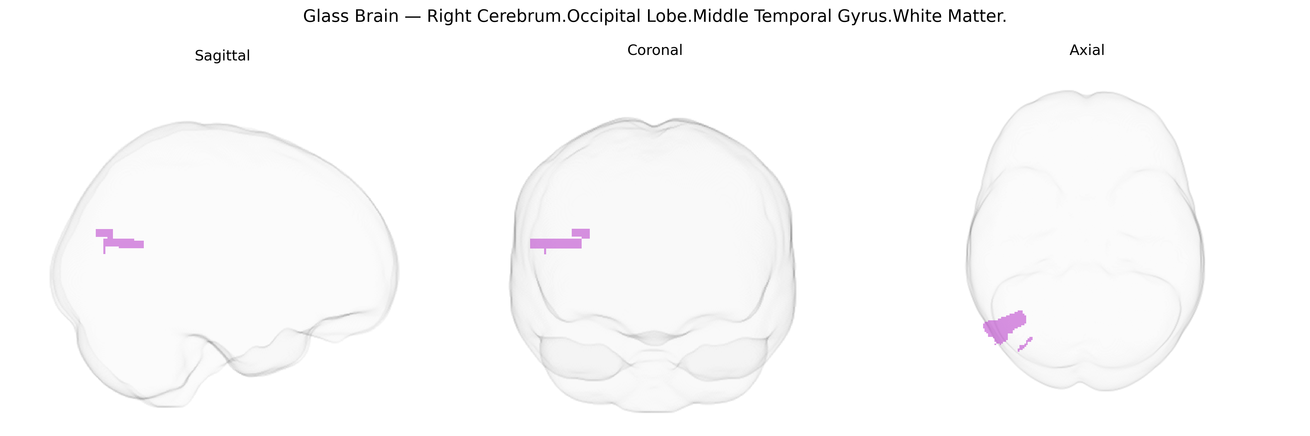
The bilateral Right Cerebrum Occipital Lobe Middle Temporal Gyrus white matter corresponds to association fiber pathways coursing beneath the middle temporal gyrus within the occipital–temporal junction of the right cerebral hemisphere. These fibers include components of the inferior longitudinal fasciculus and adjacent temporo-occipital association tracts that connect occipital visual regions with temporal cortices involved in higher-order visual processing, object recognition, and aspects of semantic integration. Functionally, this white matter supports the transfer of visual information from primary and secondary visual cortices to temporal association areas, contributing to the encoding and retrieval of complex visual features and the integration of visual input with memory and language-related processes. There is no direct link for this exact composite region; a closely related structure is the Middle temporal gyrus.
The bilateral right occipital lobe–middle temporal gyrus white matter, as defined in the Talairach 1 mm atlas, lies within posterior temporo‑occipital association pathways that have been implicated genetically through large neuroimaging GWAS of white‑matter microstructure and cortical organization rather than through region‑specific candidate studies. Polygenic influences on diffusion tensor imaging measures (e.g., fractional anisotropy and mean diffusivity) in adjacent occipito‑temporal and inferior longitudinal fasciculi have been linked to numerous loci involving axon guidance, myelination, and synaptic plasticity genes (such as variants in CNTN4, NRG1, MAG, and genes in the contactin and semaphorin families), and overlap strongly with schizophrenia, bipolar disorder, major depression, autism spectrum disorder, and ADHD polygenic risk scores, consistent with shared neurodevelopmental architectures. Occipito‑temporal white matter integrity in this general region has also been associated with genetic liability to dyslexia and other reading and language traits (e.g., KIAA0319, DCDC2, and related loci), as well as with visual processing and face/object recognition phenotypes. More broadly, multivariate brain‑wide GWAS show that common variants influencing this region’s white‑matter properties contribute to distributed factors for cognitive performance, educational attainment, and intracranial volume, suggesting that genetic effects on this tract are embedded in highly polygenic, network‑level neurodevelopmental mechanisms rather than being uniquely specific to the right occipital–middle temporal white matter alone.
Overview generated by GPT-4o (2026).
Region ID: 701
Hemisphere: bilateral
Atlas: Talairach labels 1mm
Full Quality Version: Download MP4
Full Quality Version: Download MP4
Wali Sidiqyar*, Gaurav Rudravaram*, Elyssa M. McMaster, Trent M. Schwartz, Adam M. Saunders, Kurt G. Schilling, Bennett A. Landman "Introducing SPINS: A Shared Public Visualization Library of Neuroanatomical Structures." Medical Imaging with Deep Learning- short paper
This resource is licensed under CC0 1.0 Universal (Public Domain).