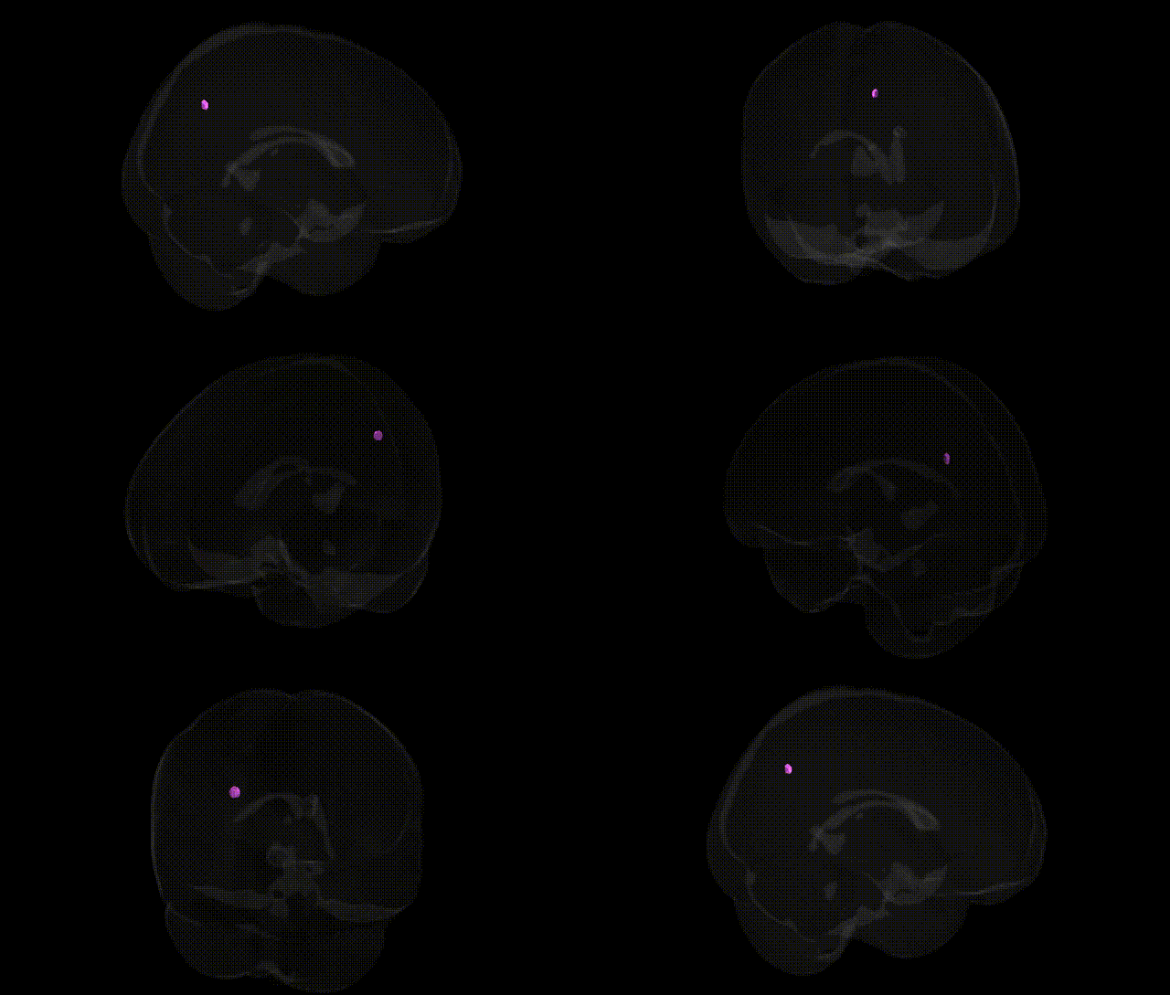
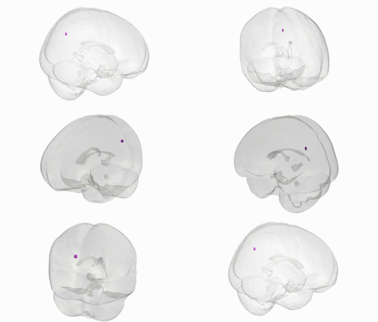
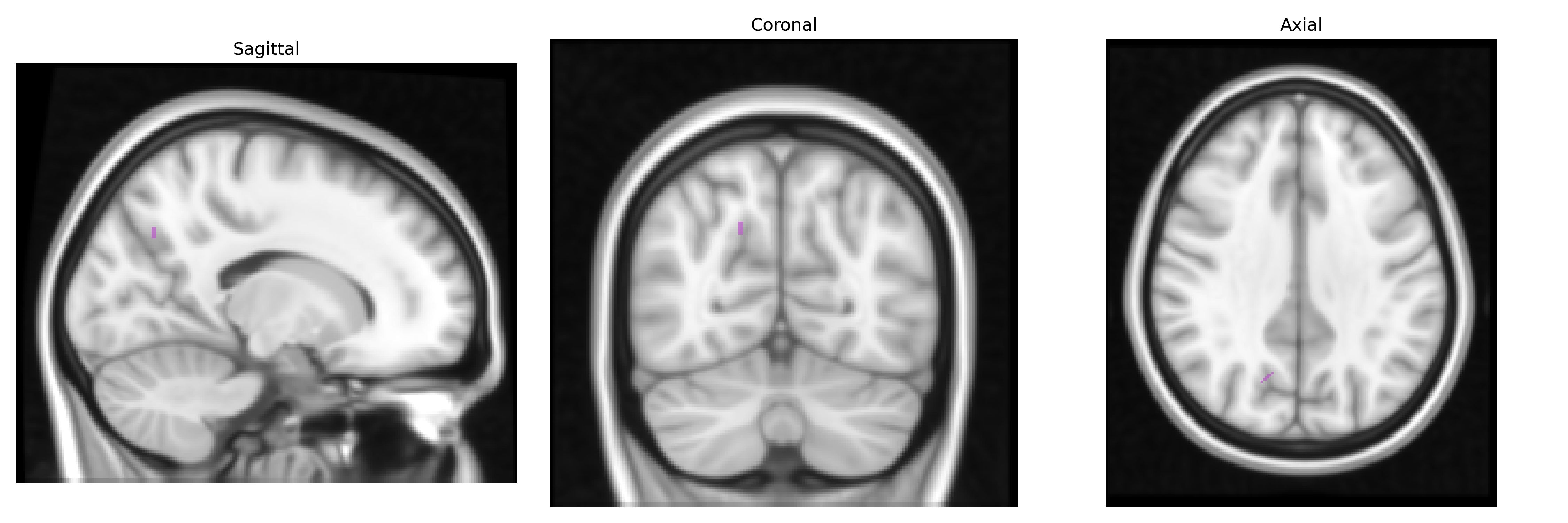
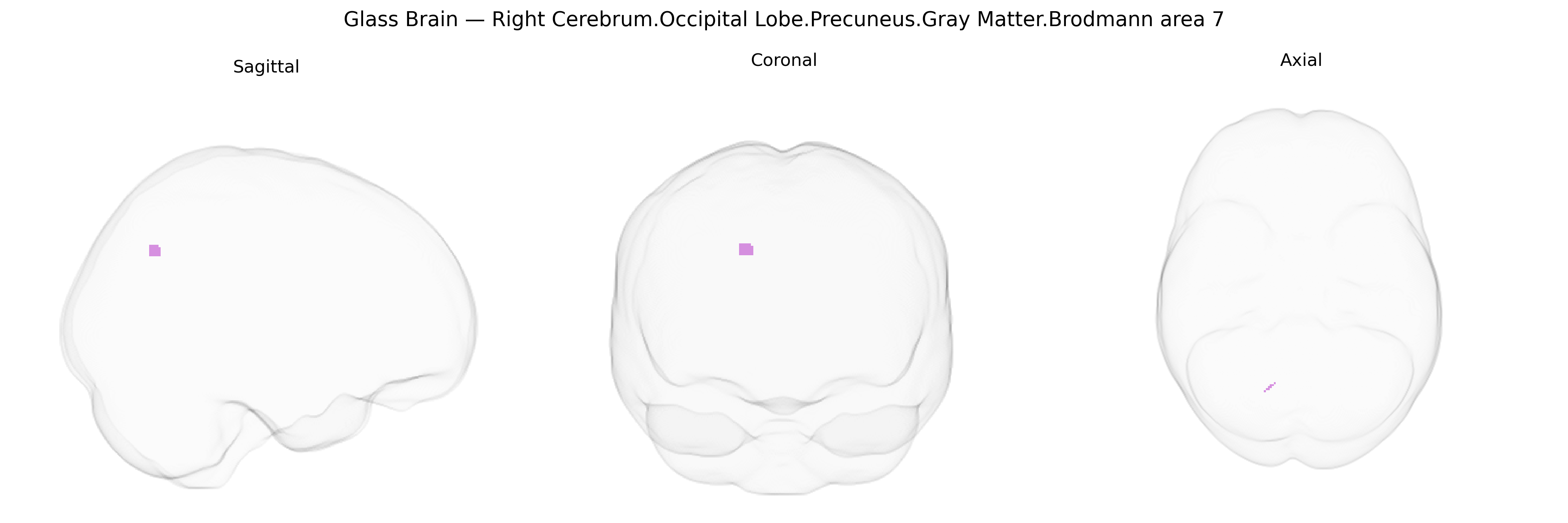
The bilateral Right Cerebrum.Occipital Lobe.Precuneus.Gray Matter.Brodmann area 7 corresponds to a segment of the superior posterior medial cortex located along the medial wall of the parietal–occipital junction, extending within the precuneus and involved primarily in higher-order visuospatial integration and sensorimotor coordination. As part of Brodmann area 7, this region participates in the transformation of visual and somatosensory information into coordinated actions, contributing to spatial attention, mental imagery, and aspects of visuomotor planning. It receives multimodal input from visual, somatosensory, and associative cortical areas and projects to premotor and other association regions, supporting complex processes such as spatial orientation, navigation, and integration of self-referential and environmental information. There is no direct Wikipedia article for this exact Talairach label; a related structure is the Precuneus.
The bilateral right precuneus gray matter in Brodmann area 7 (occipital/parietal border) has been repeatedly implicated in imaging genetics and GWAS of brain structure and function, although few variants are uniquely specific to this subregion. Large neuroimaging GWAS (e.g., ENIGMA, UK Biobank) have identified common variants in or near genes such as HMGA2, IGF1, KIAA0586, WNT3, and PAX6 that influence global and regional cortical thickness and surface area, with parietal–precuneus measures among the significantly heritable phenotypes (heritability typically ~40–70%). Polygenic architectures for Alzheimer’s disease, schizophrenia, major depression, autism spectrum disorder, and ADHD have been associated with altered precuneus morphology or connectivity, including case–control and PRS-based studies showing disease-associated thinning, volume reduction, or network dysfunction in BA7 regions; these effects are often mediated by risk loci such as APOE, CLU, BIN1, and other Alzheimer’s GWAS hits that track posterior cortical atrophy, and by schizophrenia and depression risk variants that affect default-mode network hubs including the precuneus. Cognitive GWAS on traits such as general intelligence, working memory, and visuospatial ability have linked polygenic scores to structural and functional variation in the precuneus, consistent with its role in high-level integration and self-referential processing. Epigenetic and transcriptomic work indicates that risk genes for neurodegenerative and psychiatric disorders show enriched expression or regulatory activity in association cortex including BA7, supporting a model in which highly connected hubs like the precuneus are vulnerable convergence points for diverse genetic influences on cognition and psychopathology, rather than targets of single, region-specific loci.
Overview generated by GPT-4o (2026).
Region ID: 969
Hemisphere: bilateral
Atlas: Talairach labels 1mm
Full Quality Version: Download MP4
Full Quality Version: Download MP4
Wali Sidiqyar*, Gaurav Rudravaram*, Elyssa M. McMaster, Trent M. Schwartz, Adam M. Saunders, Kurt G. Schilling, Bennett A. Landman "Introducing SPINS: A Shared Public Visualization Library of Neuroanatomical Structures." Medical Imaging with Deep Learning- short paper
This resource is licensed under CC0 1.0 Universal (Public Domain).