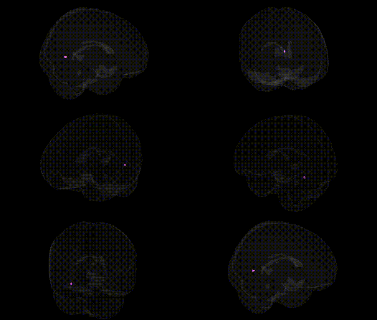
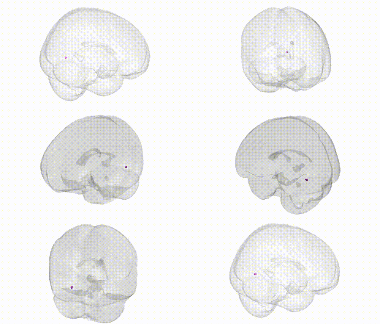
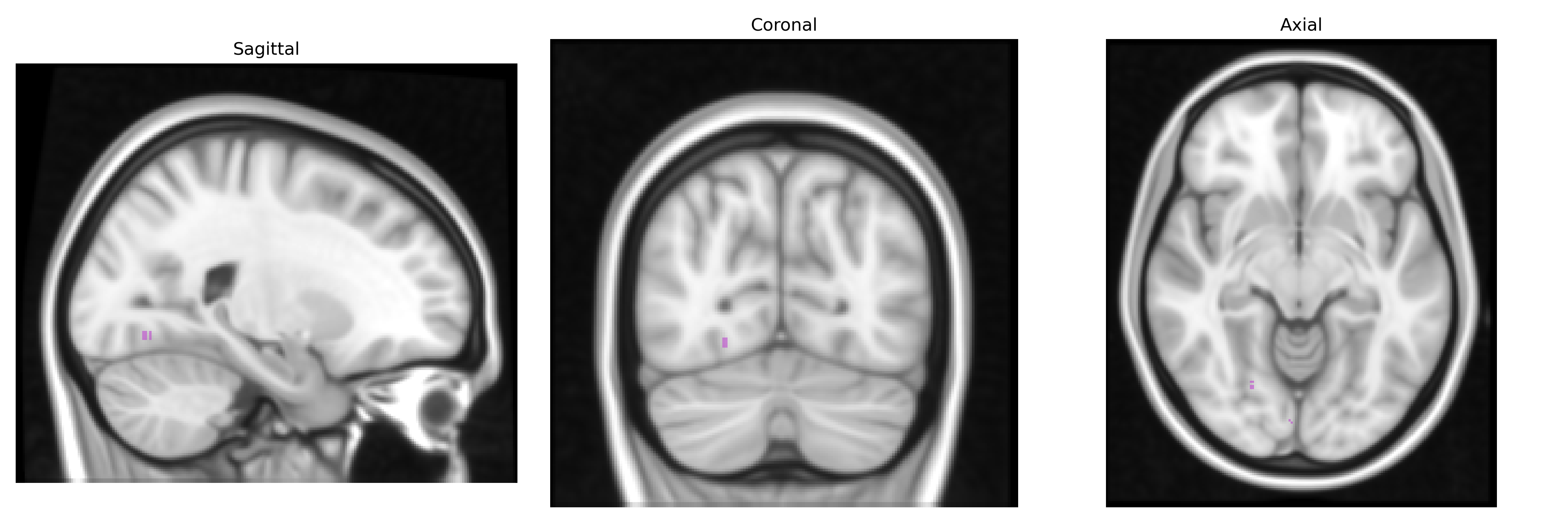
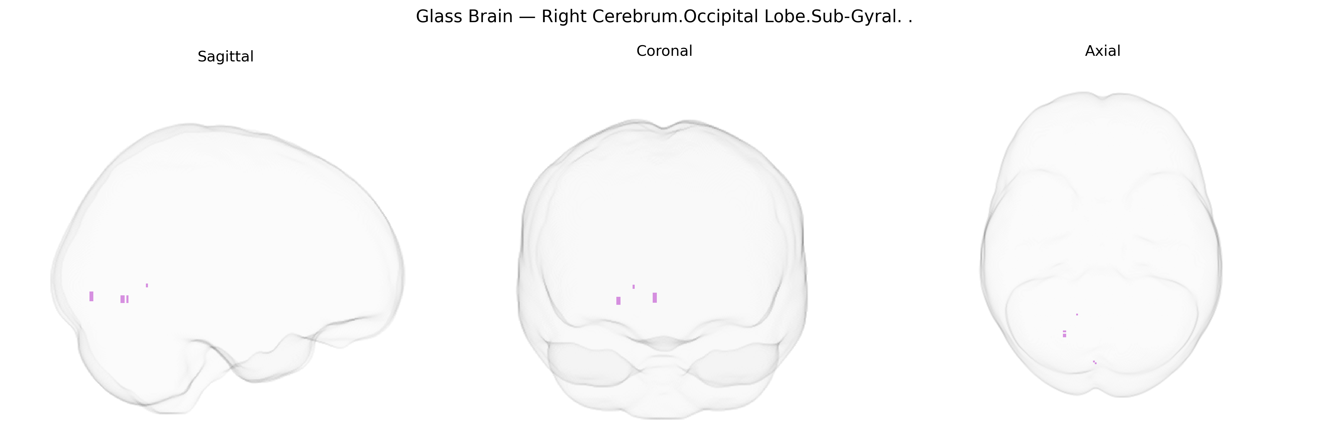
The bilateral Right Cerebrum.Occipital Lobe.Sub-Gyral region, as defined in the Talairach 1 mm atlas, refers to subcortical white matter and adjacent deep tissue lying beneath the cortical gyri of the right occipital lobe. This area primarily contains association and projection fiber tracts that connect occipital visual cortices with other cortical and subcortical regions, supporting the integration, relay, and modulation of visual information rather than direct sensory reception. Functionally, the sub-gyral occipital white matter contributes to visual processing pathways involved in perception, spatial analysis, and higher-order visual cognition by linking primary and secondary visual areas to parietal, temporal, and frontal networks. There is no direct link for the “occipital sub-gyral” region; a closely related structure is the Occipital lobe.
The bilateral Right Cerebrum Occipital Lobe Sub-Gyral region, as defined in the Talairach 1 mm atlas, corresponds mainly to deep portions of primary and associative visual cortices, and although genetic studies rarely target this exact label, multiple GWAS and imaging-genetics analyses implicate overlapping occipital and subcortical visual areas in traits and disorders with known genetic components. Large-scale brain MRI GWAS from consortia such as ENIGMA and UK Biobank have identified common variants in genes involved in neurodevelopment (e.g., HMGA2, IGF1, MAPT, and multiple loci in 3p24–p25 and 6q), associated with occipital lobe volume, cortical thickness, and surface area, while polygenic scores for general cognitive ability and educational attainment also correlate with occipital morphometry. Visual cortical and adjacent white-matter abnormalities involving occipital sub-gyral regions have been reported in genetic epilepsies (e.g., photosensitive and occipital epilepsies), neurodevelopmental disorders including autism spectrum disorder and dyslexia, and monogenic conditions affecting myelination (e.g., PLP1, SOX10, and other leukodystrophy-related genes), although these findings often implicate broader visual and posterior networks rather than this subregion alone. GWAS of migraine, especially migraine with aura, and of occipital neural traits have repeatedly implicated genes affecting vascular tone and cortical excitability (e.g., CACNA1A, PRRT2, and multiple polygenic loci) that are thought to influence occipital cortical susceptibility to spreading depolarization, consistent with functional involvement of occipital sub-gyral cortex. Additionally, schizophrenia, bipolar disorder, and major depression GWAS–linked polygenic risk scores are associated with subtle reductions in occipital gray and white matter volumes, suggesting that widespread risk variants in synaptic and neurodevelopmental genes contribute to structural variation in this region as part of distributed cortical networks rather than through region-specific genetic effects.
Overview generated by GPT-4o (2026).
Region ID: 314
Hemisphere: bilateral
Atlas: Talairach labels 1mm
Full Quality Version: Download MP4
Full Quality Version: Download MP4
Wali Sidiqyar*, Gaurav Rudravaram*, Elyssa M. McMaster, Trent M. Schwartz, Adam M. Saunders, Kurt G. Schilling, Bennett A. Landman "Introducing SPINS: A Shared Public Visualization Library of Neuroanatomical Structures." Medical Imaging with Deep Learning- short paper
This resource is licensed under CC0 1.0 Universal (Public Domain).