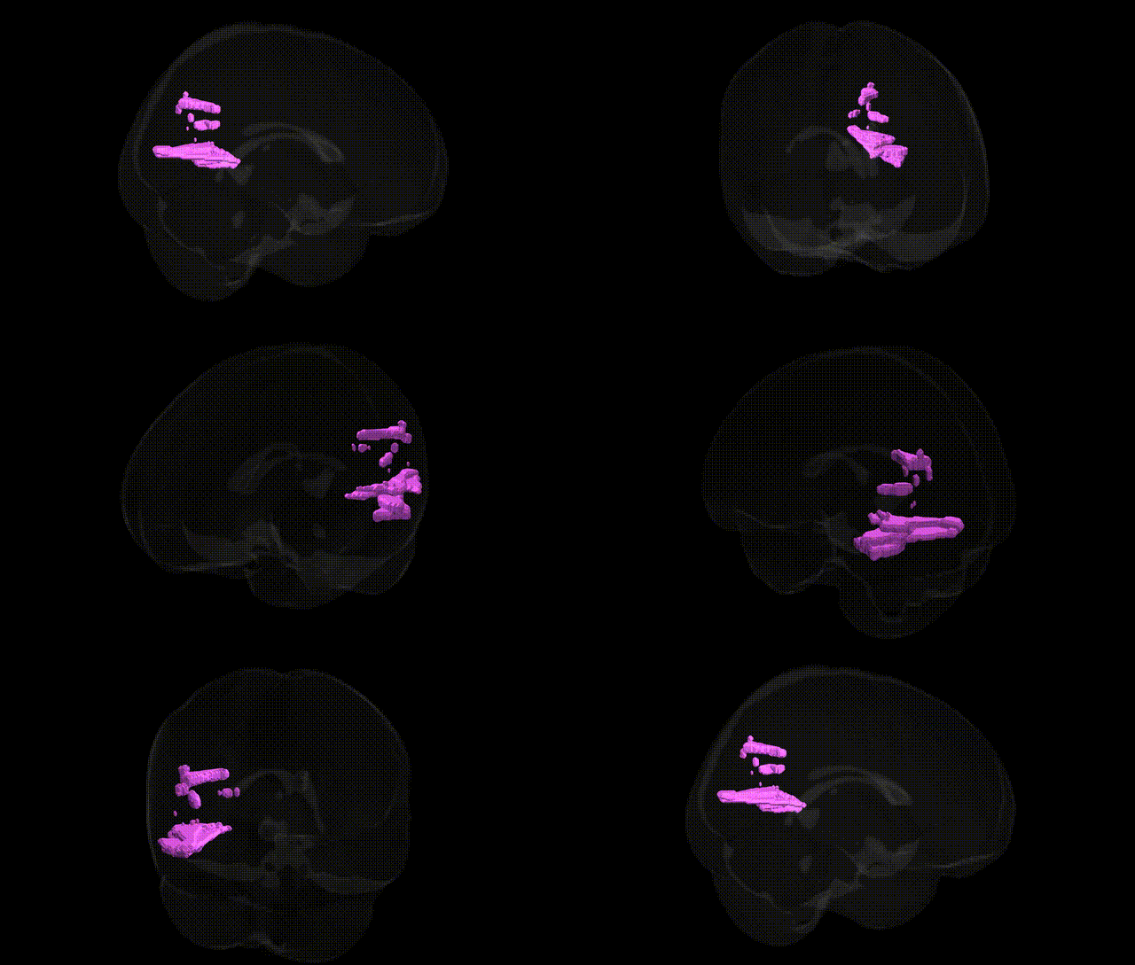
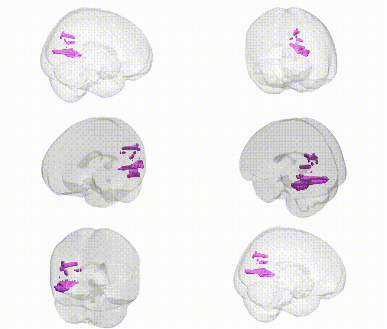
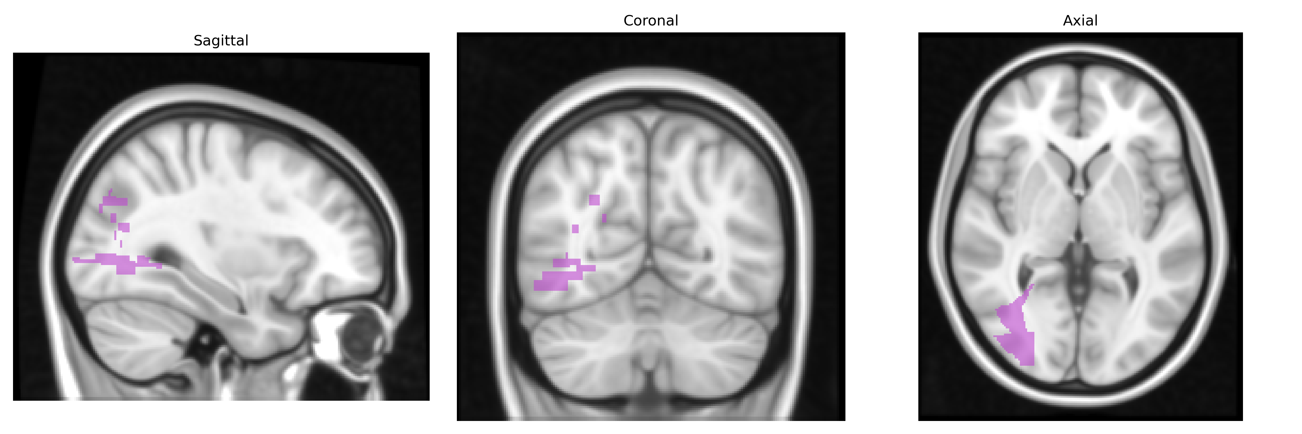
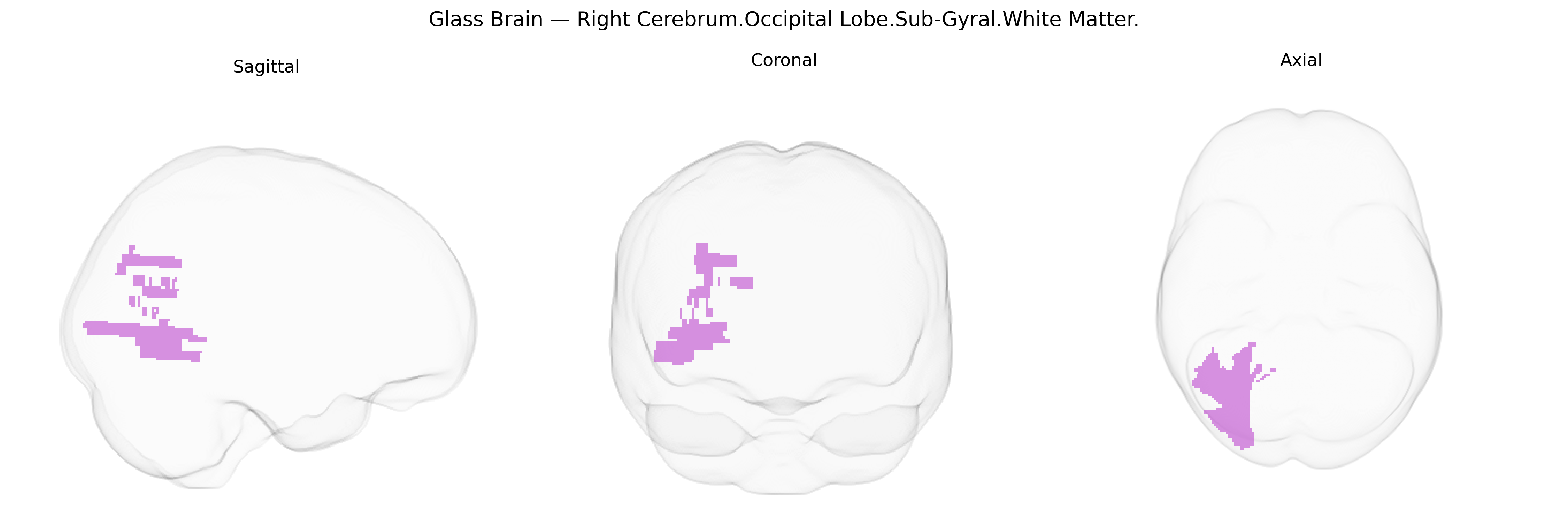
The bilateral Right Cerebrum.Occipital Lobe.Sub-Gyral.White Matter corresponds to deep white matter tracts located beneath the cortical gyri of the occipital lobe on the right side, within both hemispheres when considered bilaterally. This region contains myelinated axons that interconnect primary and associative visual cortices, and link occipital areas with parietal, temporal, and frontal regions via projection, association, and commissural fibers, thereby supporting visual perception, visuospatial processing, and higher-order visual integration. Damage or disruption to these occipital sub-gyral white matter pathways can contribute to visual field defects, impaired visual recognition, and deficits in visually guided behavior, depending on the specific tracts involved. There is no direct Wikipedia article for this exact sub-gyral white matter label; a closely related structure is the Occipital lobe.
The bilateral Right Cerebrum Occipital Lobe Sub-Gyral White Matter, as defined in the Talairach 1 mm atlas, corresponds largely to deep occipital white matter supporting visual processing networks, and genetic associations for this region arise mainly from neuroimaging GWAS of brain structure rather than region-specific candidate gene studies. Large-scale studies such as ENIGMA and UK Biobank have identified numerous common variants (e.g., in genes involved in axon guidance, myelination, and synaptic function such as CNTNAP2, NRG1, PLEKHM1, and multiple loci near oligodendrocyte-related genes) that influence occipital white matter volume, microstructure (fractional anisotropy, mean diffusivity), and cortical connectivity. These occipital white matter measures have been implicated as intermediate phenotypes in neurodevelopmental and psychiatric disorders—including schizophrenia, major depression, bipolar disorder, autism spectrum disorder, ADHD, and dyslexia—where polygenic risk scores for these conditions correlate with alterations in posterior white matter tracts (e.g., inferior longitudinal fasciculus, optic radiations) coursing through the occipital sub-gyral region. Additionally, GWAS of visual processing and reading-related traits have linked variants in genes such as KIAA0319, DCDC2, and ROBO1 to structural and connectivity differences in posterior occipito-temporal and occipital white matter, although these findings are typically reported at the tract or lobar level rather than at the fine-grained Talairach label used here. Overall, genetic influences on this region appear highly polygenic, reflecting general mechanisms of white matter development and maintenance, with its involvement in visual and higher-order cognitive networks contributing to observed associations with a range of neurodevelopmental, psychiatric, and cognitive traits.
Overview generated by GPT-4o (2026).
Region ID: 320
Hemisphere: bilateral
Atlas: Talairach labels 1mm
Full Quality Version: Download MP4
Full Quality Version: Download MP4
Wali Sidiqyar*, Gaurav Rudravaram*, Elyssa M. McMaster, Trent M. Schwartz, Adam M. Saunders, Kurt G. Schilling, Bennett A. Landman "Introducing SPINS: A Shared Public Visualization Library of Neuroanatomical Structures." Medical Imaging with Deep Learning- short paper
This resource is licensed under CC0 1.0 Universal (Public Domain).