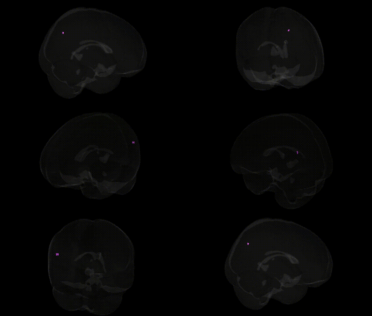
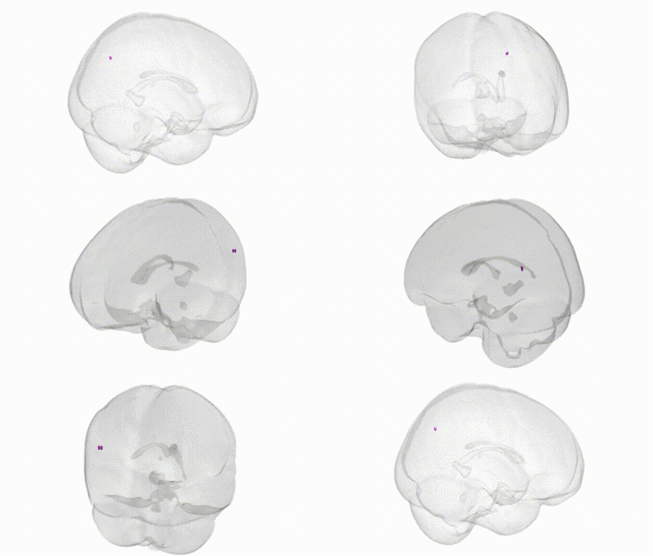
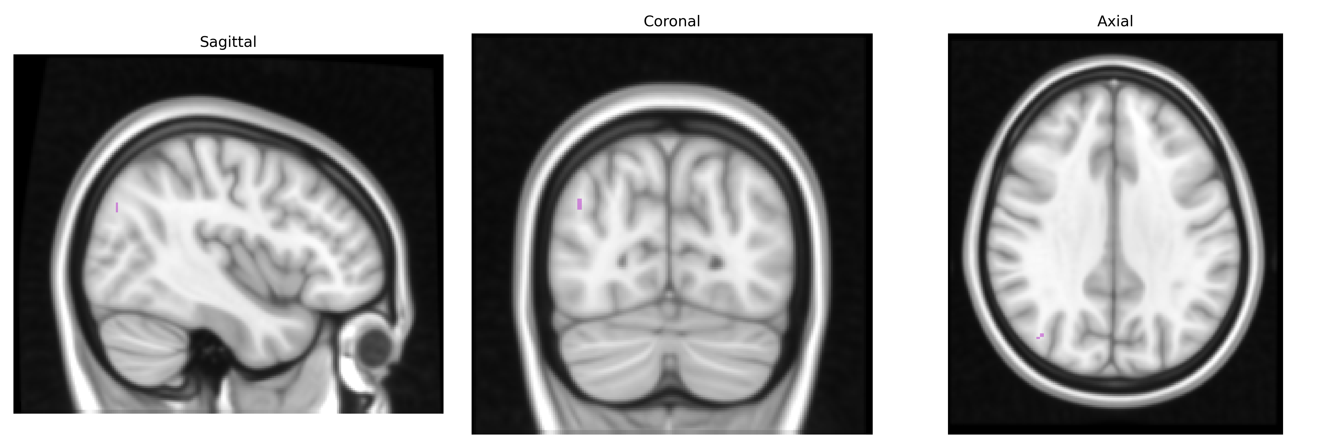
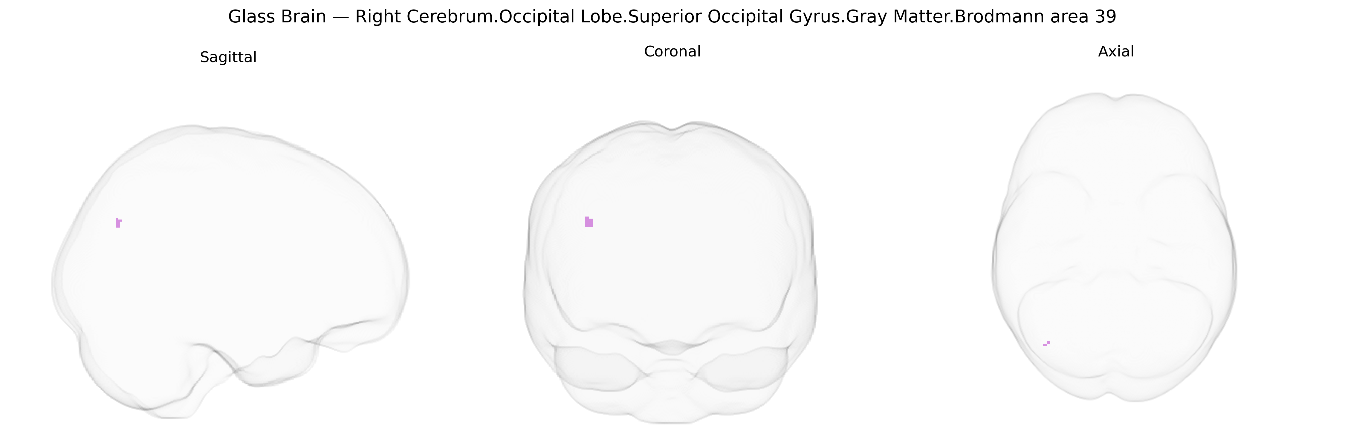
The bilateral Right Cerebrum.Occipital Lobe.Superior Occipital Gyrus.Gray Matter.Brodmann area 39 corresponds primarily to the angular gyrus region situated at the junction of the occipital, parietal, and temporal lobes, within the inferior parietal lobule bordering posterior occipital cortex in the Talairach 1 mm atlas. This heteromodal association cortex integrates visual, auditory, and somatosensory information and is heavily implicated in language-related functions (reading, writing, semantic processing), visuospatial cognition, number processing, and aspects of attention and theory of mind. Anatomically, it receives rich input from primary and secondary visual cortices and from temporal association areas, and projects to widespread frontoparietal networks, making it a key hub in the default mode and language networks. Functionally, lesions in this region are classically associated with Gerstmann syndrome (including acalculia, agraphia, finger agnosia, and left–right disorientation) and other higher-order cognitive deficits reflecting its role in multimodal integration and symbolic processing. Brodmann area 39
The bilateral right superior occipital gyrus gray matter in Brodmann area 39 (classically corresponding mainly to the angular gyrus in the inferior parietal/temporoparietal junction rather than “occipital” cortex per se) has been repeatedly implicated in genetic studies of higher cognitive function, language, and neuropsychiatric risk through both GWAS of brain structure and imaging–genetics analyses. Large neuroimaging GWAS consortia (e.g., ENIGMA, UK Biobank) have identified common variants in genes such as HMGA2, IGF1, KIAA0586, and multiple loci in intronic or intergenic regions that influence regional cortical thickness and surface area in temporoparietal territories including BA39, with downstream associations to general intelligence, educational attainment, and reading/language skills. Polygenic scores for schizophrenia, bipolar disorder, and major depressive disorder have been linked to gray matter volume reductions and altered activation in BA39/angular gyrus regions, and risk variants in genes such as CACNA1C, ZNF804A, and GRM3 show convergent effects on functional connectivity involving BA39 during working memory and language tasks. FOXP2-related and DCDC2/KIAA0319-related language and dyslexia risk alleles have been associated with altered activation and structure within the left-lateralized BA39 region, with some bilateral effects reported in multivariate imaging–genetics models. Alzheimer’s disease GWAS hits (e.g., APOE, CLU, BIN1) and polygenic risk scores correlate with atrophy and hypometabolism in temporoparietal regions encompassing BA39, particularly in early-stage and prodromal disease, and similar patterns have been noted for frontotemporal dementia risk loci in select studies. In autism spectrum disorder and social cognition GWAS and imaging–genetics work, BA39 has emerged as part of genetically influenced “social brain” networks, with common variation in synaptic and neurodevelopmental genes (e.g., NRXN1, CNTNAP2, SHANK3 pathways) associated with atypical structure and connectivity of this region. Overall, while specific single-gene–to–BA39 mappings remain limited, convergent GWAS and polygenic analyses support BA39/superior occipital–parietal territory as a genetically modulated hub for language, higher cognition, and vulnerability to major neuropsychiatric and neurodegenerative disorders.
Overview generated by GPT-4o (2026).
Region ID: 910
Hemisphere: bilateral
Atlas: Talairach labels 1mm
Full Quality Version: Download MP4
Full Quality Version: Download MP4
Wali Sidiqyar*, Gaurav Rudravaram*, Elyssa M. McMaster, Trent M. Schwartz, Adam M. Saunders, Kurt G. Schilling, Bennett A. Landman "Introducing SPINS: A Shared Public Visualization Library of Neuroanatomical Structures." Medical Imaging with Deep Learning- short paper
This resource is licensed under CC0 1.0 Universal (Public Domain).