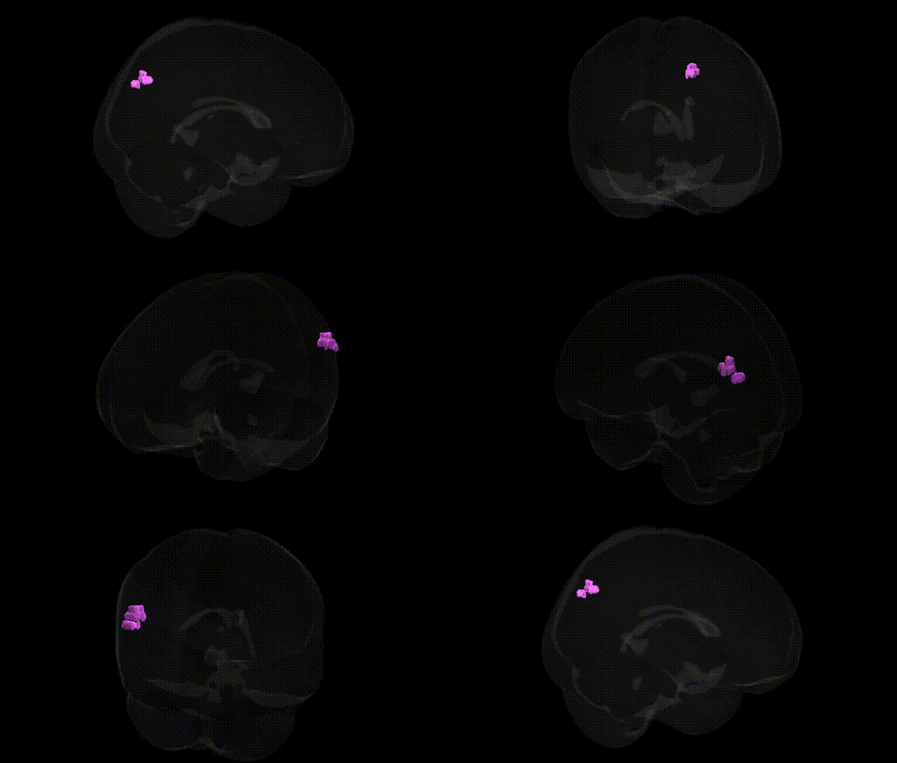
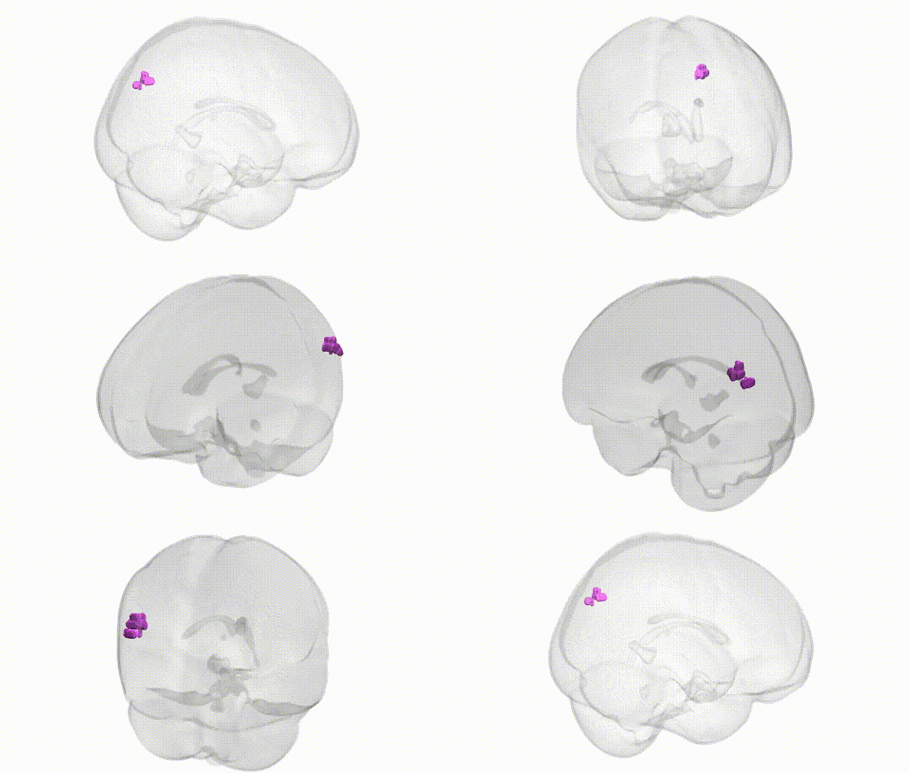
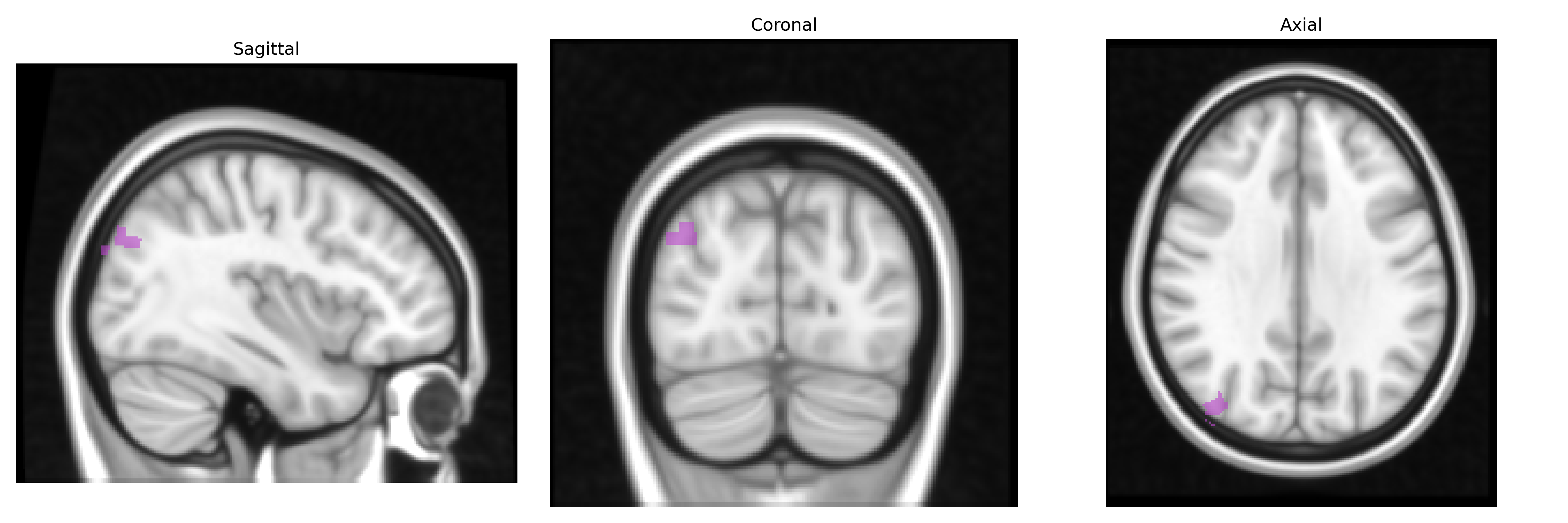
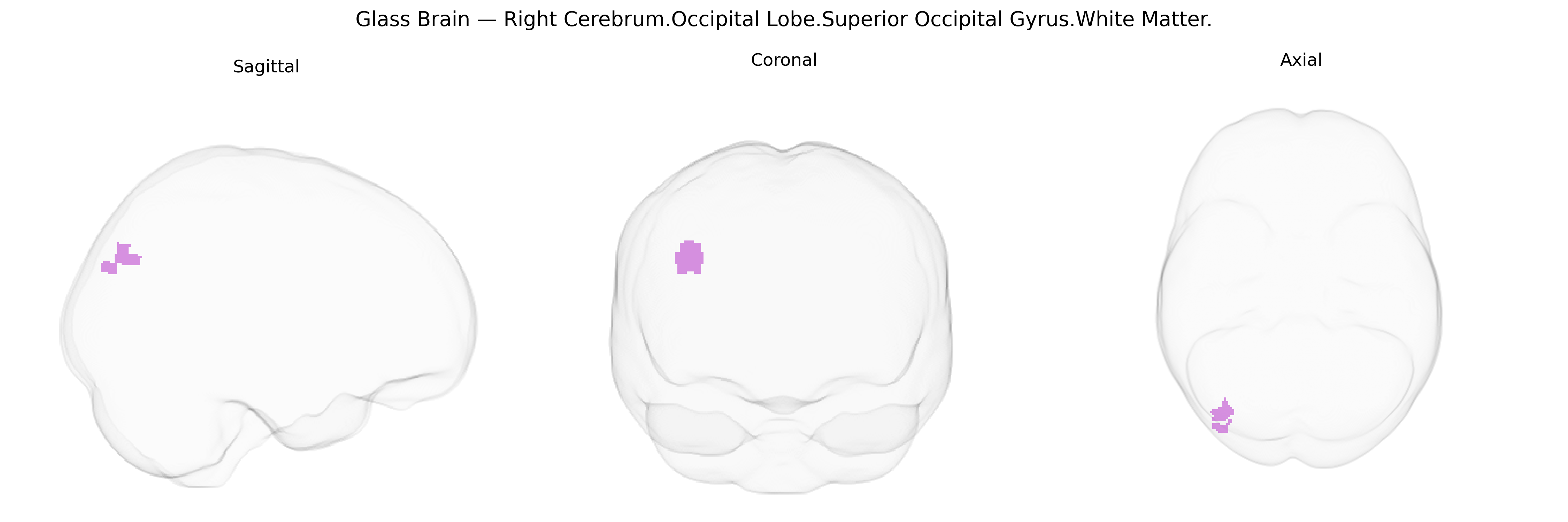
The bilateral Right Cerebrum.Occipital Lobe.Superior Occipital Gyrus.White Matter corresponds to the deep myelinated fiber pathways underlying the superior occipital gyrus in the right occipital lobe, as defined in the Talairach 1 mm atlas. These fibers include association tracts that interconnect dorsal occipital visual areas with parietal and temporal regions, contributing to the dorsal (“where/how”) visual stream involved in visuospatial processing, motion perception, and visually guided actions. The white matter here carries output from primary and secondary visual cortices toward higher-order multimodal areas, supporting integration of visual information with attention, eye movements, and spatial orientation. There is no direct link for this exact white-matter subdivision; see the related structure Occipital lobe.
The bilateral right superior occipital gyrus white matter, as defined in the Talairach 1 mm atlas, lies within posterior association pathways (including segments of the superior longitudinal and occipital callosal fibers) that have been implicated in multiple genetic and genome‑wide association studies through imaging‑genetics and diffusion MRI approaches, rather than through region‑specific GWAS alone. Polygenic influences involving genes related to myelination, axonal guidance, and synaptic plasticity—such as variants in NRG1/ERBB signaling, MAG, PLP1, and genes in the netrin and semaphorin families—have been repeatedly associated with interindividual differences in occipital and parietal white matter microstructure (fractional anisotropy and mean diffusivity) and with global white matter integrity factors that load on posterior tracts. Large‑scale imaging GWAS consortia (e.g., ENIGMA, UK Biobank) have reported that common variants in genes affecting neurodevelopment (e.g., BDNF, CNTNAP2, DISC1, and multiple 3p21, 6q22–q23, and 17q loci) show associations with diffusion metrics in superior occipital and adjacent dorsal visual stream pathways, which in turn are linked to cognitive traits such as processing speed, visuospatial ability, and general intelligence. Clinically, risk variants for schizophrenia, bipolar disorder, major depression, and autism spectrum disorders have been associated with altered integrity of dorsal occipital–parietal white matter, including superior occipital gyrus connections, consistent with findings of reduced fractional anisotropy and disrupted connectivity in these pathways; additional associations have been observed with neurodevelopmental traits (e.g., ADHD), migraine, and normal‑range variation in visual and attentional performance, though most findings implicate broader posterior white matter networks rather than this gyrus‑specific white matter alone.
Overview generated by GPT-4o (2026).
Region ID: 851
Hemisphere: bilateral
Atlas: Talairach labels 1mm
Full Quality Version: Download MP4
Full Quality Version: Download MP4
Wali Sidiqyar*, Gaurav Rudravaram*, Elyssa M. McMaster, Trent M. Schwartz, Adam M. Saunders, Kurt G. Schilling, Bennett A. Landman "Introducing SPINS: A Shared Public Visualization Library of Neuroanatomical Structures." Medical Imaging with Deep Learning- short paper
This resource is licensed under CC0 1.0 Universal (Public Domain).