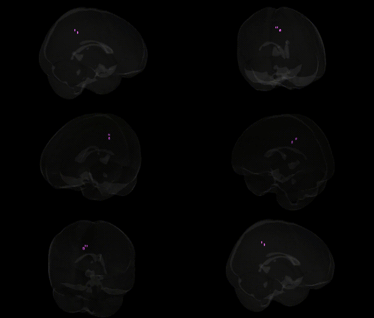
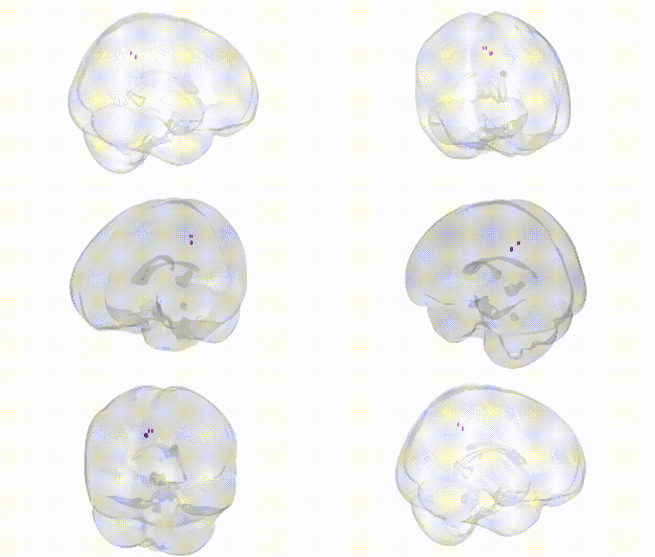
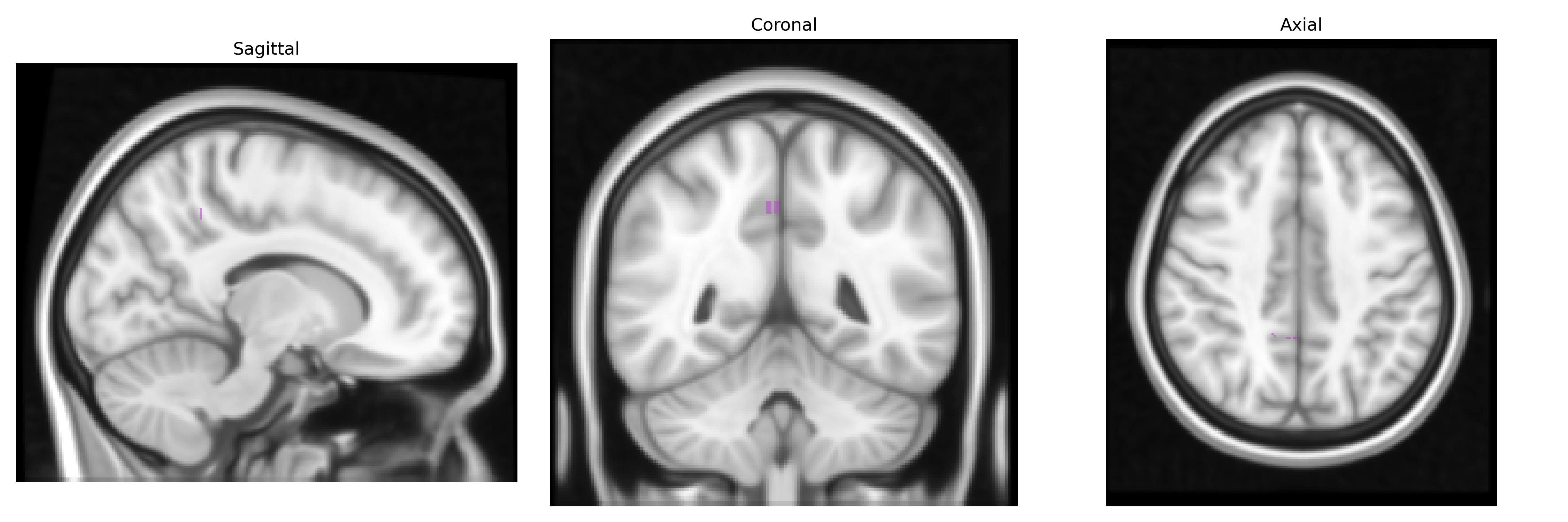
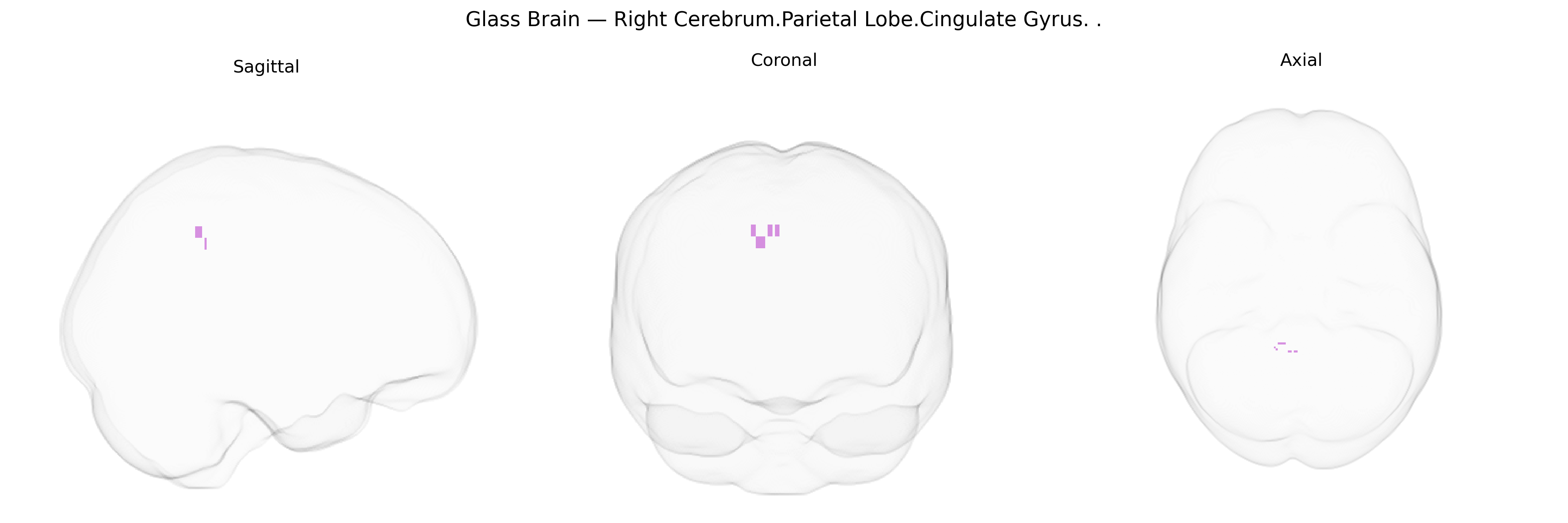
The bilateral Right Cerebrum.Parietal Lobe.Cingulate Gyrus corresponds to portions of the cingulate cortex that extend along the medial surface of the parietal lobe in both hemispheres, lying superior to the corpus callosum and posterior to the frontal cingulate regions. Functionally, this territory is often associated with the posterior cingulate cortex and adjacent medial parietal areas, which participate in internally directed cognition, visuospatial orientation, and integration of sensory and contextual information, as well as in default mode network activity related to memory and self-referential processing. Although the labeling specifies “Right Cerebrum” and “Parietal Lobe,” the bilateral designation highlights homologous regions in both hemispheres that support coordinated higher-order associative and attentional functions within the cingulate-parietal network. There is no direct link for this exact composite Talairach label; a closely related structure is the Cingulate gyrus.
The bilateral Right Cerebrum.Parietal Lobe.Cingulate Gyrus (as approximated by Talairach 1 mm labeling, spanning posterior cingulate and adjacent parietal–cingulate transition zones) is part of the default mode and attentional networks and has been repeatedly implicated in large-scale imaging genetics and GWAS of brain structure and function; variants in and near genes such as APOE, CLU, PICALM, and BIN1 show robust associations with cortical thinning, metabolic disruption, and connectivity alterations in posterior cingulate/parietal cingulate regions in Alzheimer’s disease, while polygenic risk scores for Alzheimer’s and related dementias strongly predict atrophy and hypometabolism in this area. Common variants in genes involved in synaptic plasticity, glutamatergic signaling, and myelination (e.g., BDNF, GRIN2B, NRG1, and oligodendrocyte-related loci) have been linked to individual differences in cingulate–parietal cortical thickness, surface area, and functional coupling in GWAS of brain MRI phenotypes, with downstream associations to memory, attentional control, and default mode activity. Psychiatric genetics studies show that schizophrenia, bipolar disorder, major depression, and ADHD polygenic risk scores are associated with altered structure and activation of the posterior and dorsal cingulate/parietal cingulate regions, with specific loci (such as CACNA1C, ZNF804A, and MHC-region variants) contributing to connectivity changes within cingulo-parietal and frontoparietal control networks. Additionally, imaging GWAS of traits such as neuroticism, educational attainment, and general cognitive ability implicate variants across distributed loci (including MAPT, genes involved in axonal guidance, and lipid metabolism) that modulate gray matter volume and functional connectivity in this cingulate–parietal territory, underscoring its role as a genetically influenced hub linking cognition, affect, and neurodegenerative vulnerability.
Overview generated by GPT-4o (2026).
Region ID: 997
Hemisphere: bilateral
Atlas: Talairach labels 1mm
Full Quality Version: Download MP4
Full Quality Version: Download MP4
Wali Sidiqyar*, Gaurav Rudravaram*, Elyssa M. McMaster, Trent M. Schwartz, Adam M. Saunders, Kurt G. Schilling, Bennett A. Landman "Introducing SPINS: A Shared Public Visualization Library of Neuroanatomical Structures." Medical Imaging with Deep Learning- short paper
This resource is licensed under CC0 1.0 Universal (Public Domain).