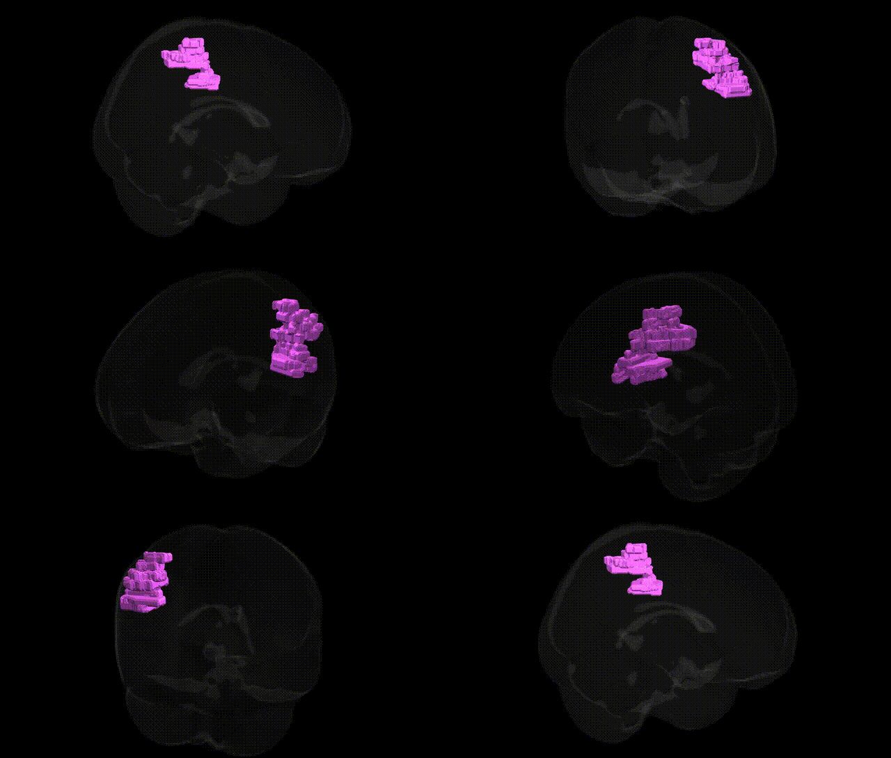
The bilateral Right Cerebrum Parietal Lobe Inferior Parietal Lobule White Matter corresponds to the deep myelinated fiber tracts underlying the inferior parietal lobule (including the supramarginal and angular gyri) in the right cerebral hemisphere. This white matter region participates in cortico-cortical and cortico-subcortical connectivity supporting multimodal integration of visual, auditory, and somatosensory information, and contributes to higher cognitive functions such as language, spatial attention, number processing, and aspects of social cognition. Fibers here are part of major association pathways (e.g., components of the superior longitudinal fasciculus and related tracts) that link the inferior parietal cortex with frontal and temporal regions. There is no direct Wikipedia article for the white matter of the inferior parietal lobule; a closely related cortical structure is the Inferior parietal lobule.
The bilateral right inferior parietal lobule (IPL) white matter, as defined in the Talairach 1 mm atlas, has been implicated in several genetic and genome‑wide association findings through its roles in higher‑order cognition, language, and attention. GWAS and imaging‑genetics studies of white‑matter microstructure (e.g., fractional anisotropy and mean diffusivity) repeatedly implicate variants affecting axon guidance, myelination, and synaptic function—such as common variants in genes including CNTNAP2, NRG1/NRG3, RELN, and those within major neurodevelopmental pathways—in modulating structural integrity of parietal association tracts that traverse the IPL, particularly components of the superior longitudinal fasciculus and arcuate system. Polygenic risk for schizophrenia, autism spectrum disorder, and attention‑deficit/hyperactivity disorder shows robust associations with altered right parietal white‑matter metrics, and risk loci in genes such as CACNA1C, ZNF804A, and GRIN2A have been linked to disrupted frontoparietal and temporoparietal connectivity involving the IPL. Large GWAS of brain morphometry (e.g., ENIGMA, UK Biobank) report heritable variation in parietal lobe volume and surface area, with loci in genes related to neurodevelopment (e.g., HMGA2, IGF1, and Wnt pathway components) influencing association cortex size that encompasses the IPL. Cognitive and educational attainment GWAS, whose polygenic scores correlate with right IPL structure and connectivity, implicate genes involved in synaptic plasticity (e.g., BDNF pathway, NMDA receptor subunits) that are enriched in parietal association networks. Additionally, genetic risk for Alzheimer’s disease and other dementias (including APOE and CLU variants) has been associated with early microstructural changes and hypometabolism in parietal association regions, including the inferior parietal lobule white matter, consistent with this region’s vulnerability in neurodegenerative and neuropsychiatric disorders.
Overview generated by GPT-4o (2026).
Region ID: 880
Hemisphere: bilateral
Atlas: Talairach labels 1mm
Full Quality Version: Download MP4
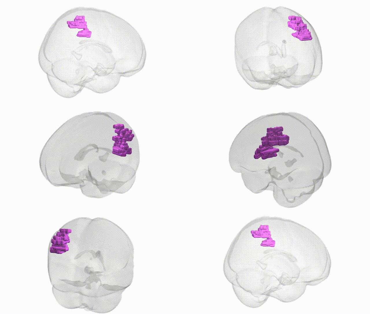
Full Quality Version: Download MP4
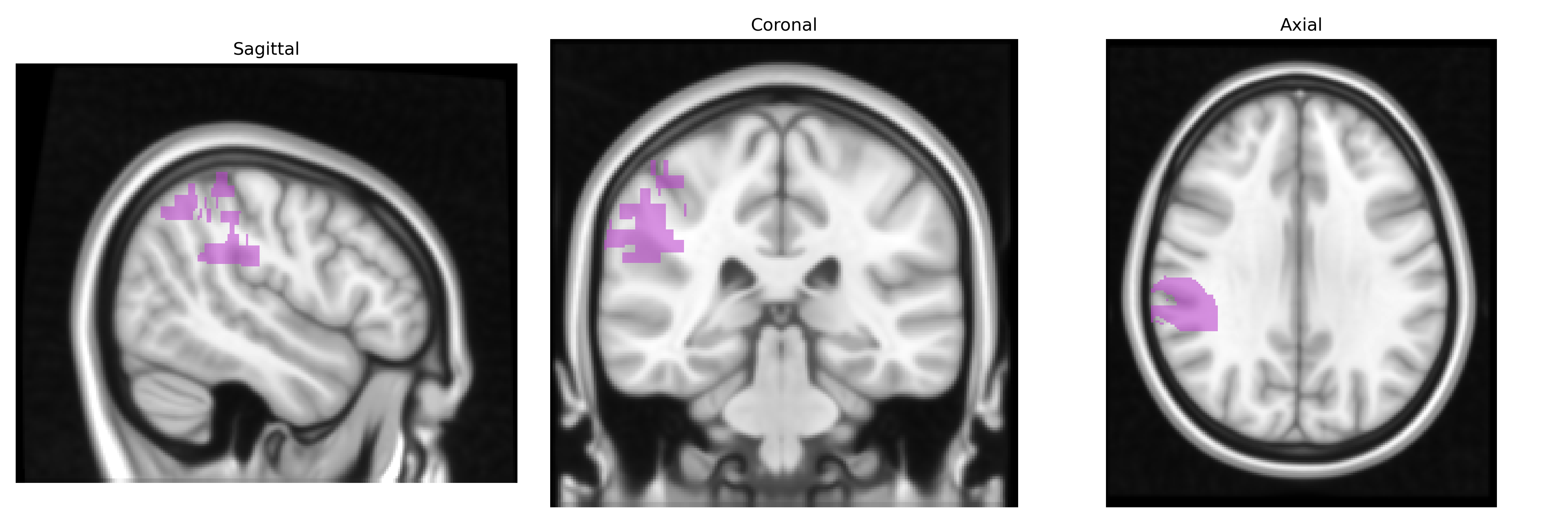
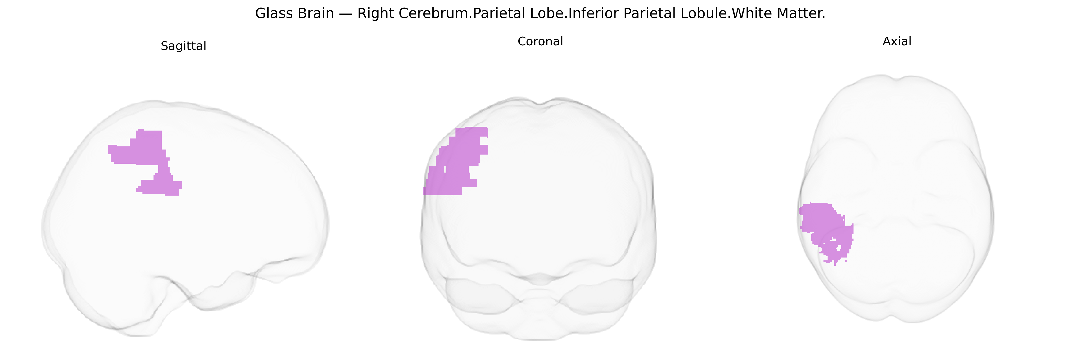
Wali Sidiqyar*, Gaurav Rudravaram*, Elyssa M. McMaster, Trent M. Schwartz, Adam M. Saunders, Kurt G. Schilling, Bennett A. Landman "Introducing SPINS: A Shared Public Visualization Library of Neuroanatomical Structures." Medical Imaging with Deep Learning- short paper
This resource is licensed under CC0 1.0 Universal (Public Domain).