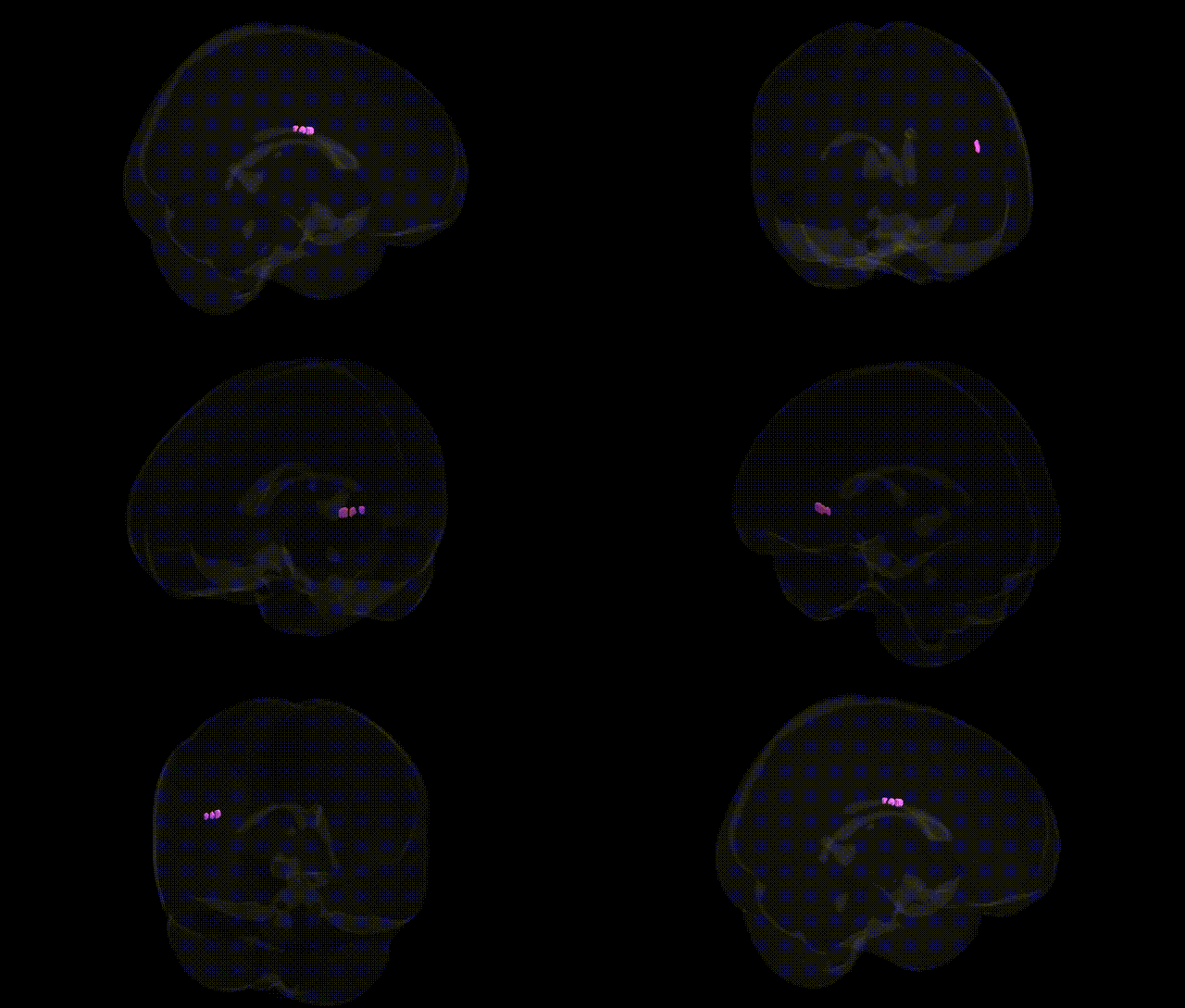
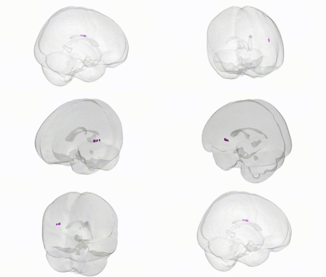
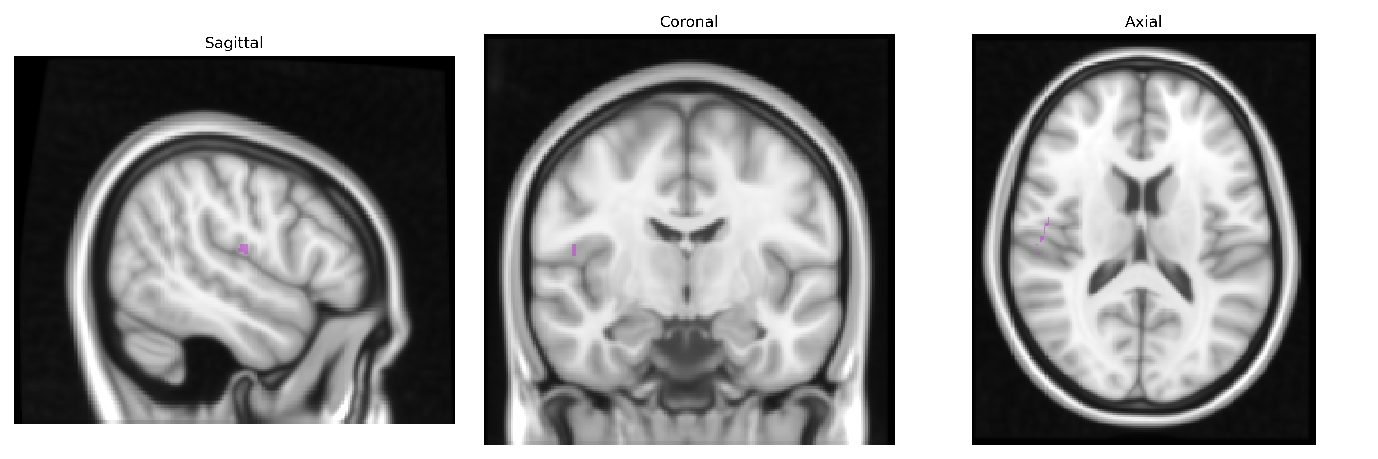
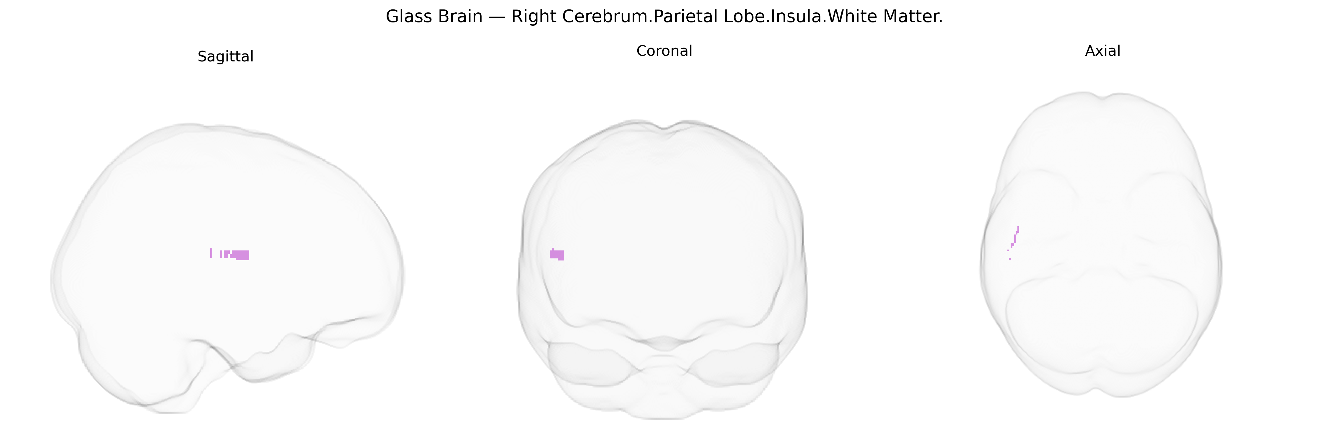
The bilateral Right Cerebrum.Parietal Lobe.Insula.White Matter region, as defined in the Talairach 1 mm atlas, corresponds to subcortical white matter tracts situated deep to the insular cortex but topographically grouped with the parietal lobe. This white matter comprises association and projection fibers that interconnect the insula—an area involved in interoception, multimodal sensory integration, affective processing, and autonomic regulation—with parietal, frontal, and temporal cortical regions, as well as subcortical structures. Functionally, these pathways contribute to integration of somatosensory, visceral, and emotional information, and support higher-order processes such as body awareness, pain perception, and aspects of language and attention. There is no direct Wikipedia article for this exact white matter label; a related cortical structure is the Insular cortex.
The bilateral Right Cerebrum.Parietal Lobe.Insula.White Matter, as defined in the Talairach 1 mm atlas, encompasses association fiber pathways linking parietal and insular cortices that participate in somatosensory integration, interoception, vestibular processing, and salience detection, and genetic associations are typically inferred from GWAS of white-matter microstructure (e.g., diffusion tensor MRI) rather than this label specifically. Large imaging-genetics consortia such as ENIGMA and UK Biobank have shown that white-matter integrity in parietal–insular and adjacent association tracts (including superior longitudinal and arcuate fasciculi and short U-fibers) is highly heritable and influenced by common variants in genes involved in axon guidance, myelination, and oligodendrocyte function (for example, NRG1/ERBB signaling genes, MAG, MBP, LINGO1, and loci near NCAN and CNTN4), as well as broader neurodevelopmental loci such as those in the major histocompatibility complex. GWAS of diffusion metrics have reported associations between parietal and insular white-matter measures and polygenic risk for schizophrenia, bipolar disorder, major depression, and autism, consistent with case–control findings of altered parietal–insular connectivity in these disorders, while polygenic scores for cognitive ability and educational attainment also correlate with higher fractional anisotropy in overlapping parietal association tracts. Additional links have been described between variants in myelin- and lipid-related genes and susceptibility to small-vessel disease and leukoaraiosis affecting parietal and perisylvian white matter, and between migraine or pain-related loci (e.g., near TRPM8 and other nociceptive or vascular genes) and structural or functional alterations in insular–parietal networks implicated in pain and interoceptive processing. Overall, genetic findings converge on pathways regulating neurodevelopment, synaptic plasticity, and white-matter integrity, with pleiotropic effects across psychiatric, cognitive, and vascular phenotypes that are partially mediated through structural variation in parietal–insular white matter, even though the exact Talairach-defined region is not typically isolated as a unique GWAS target.
Overview generated by GPT-4o (2026).
Region ID: 777
Hemisphere: bilateral
Atlas: Talairach labels 1mm
Full Quality Version: Download MP4
Full Quality Version: Download MP4
Wali Sidiqyar*, Gaurav Rudravaram*, Elyssa M. McMaster, Trent M. Schwartz, Adam M. Saunders, Kurt G. Schilling, Bennett A. Landman "Introducing SPINS: A Shared Public Visualization Library of Neuroanatomical Structures." Medical Imaging with Deep Learning- short paper
This resource is licensed under CC0 1.0 Universal (Public Domain).