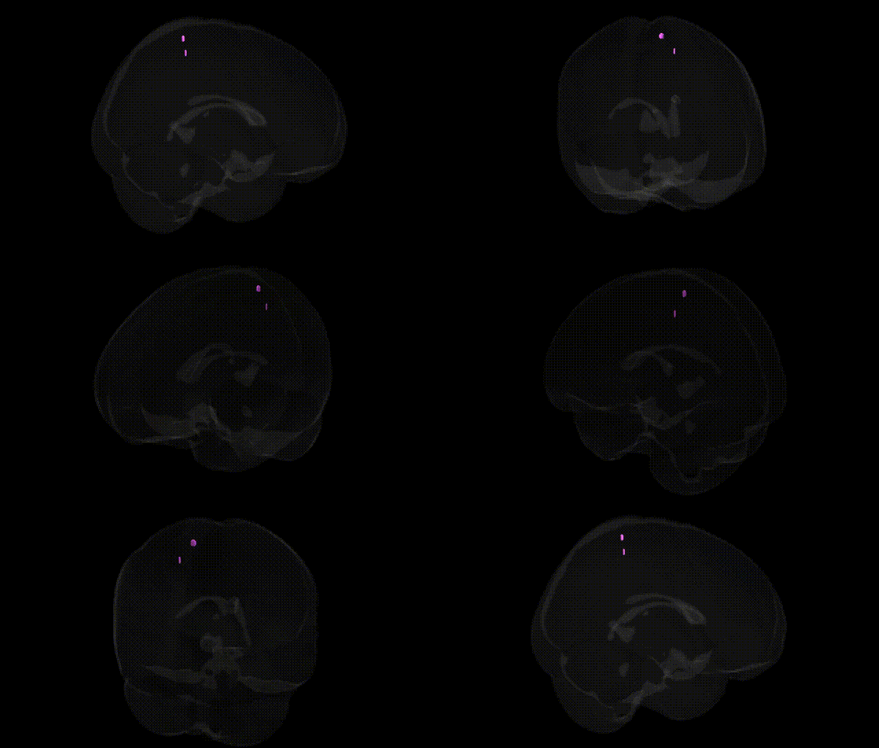
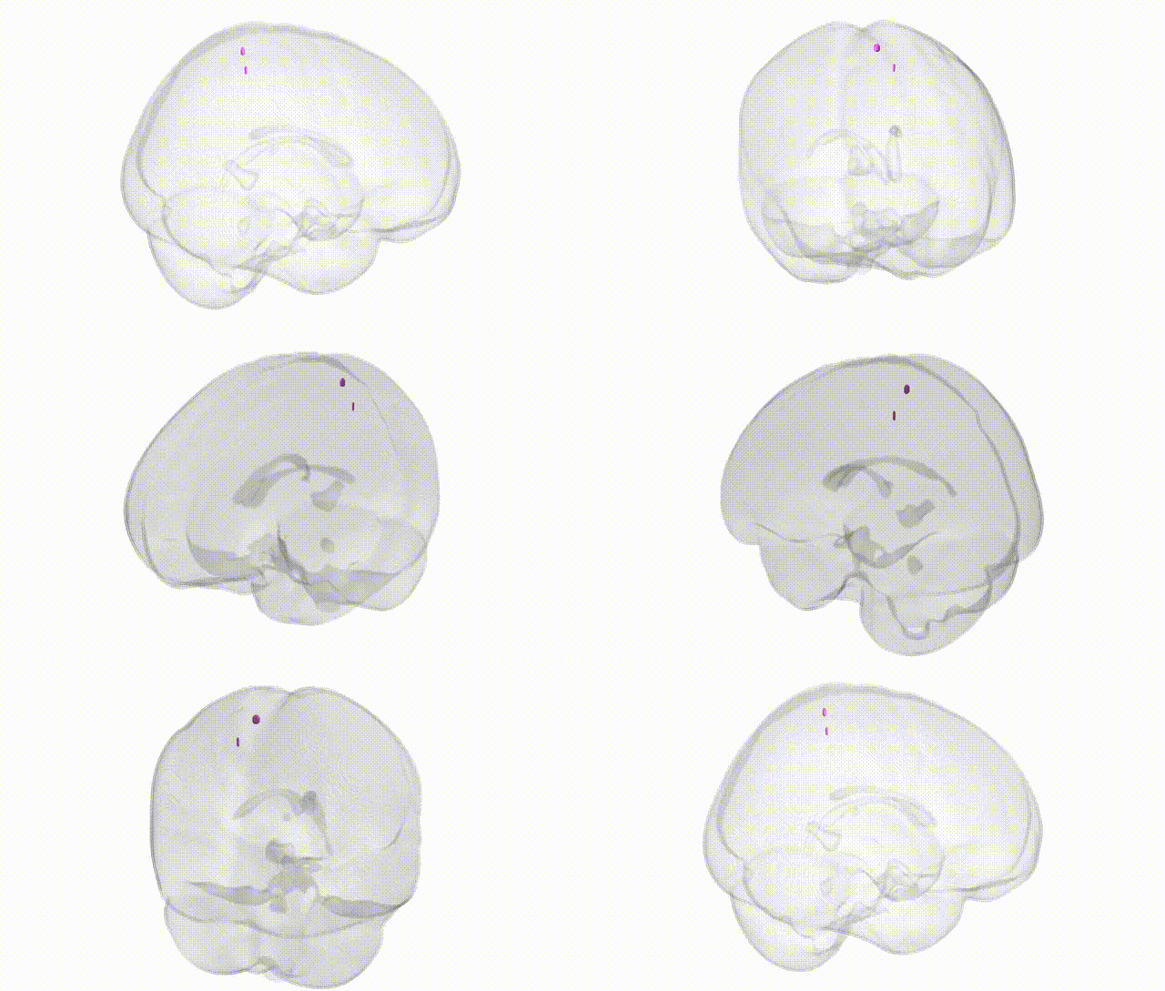
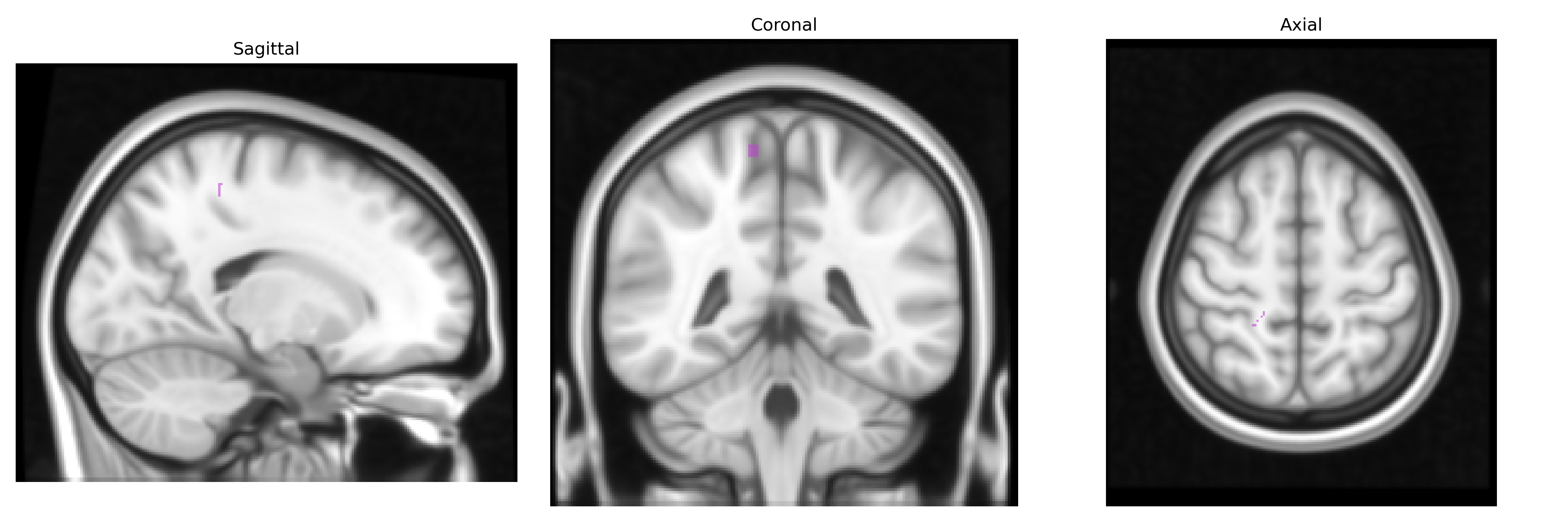
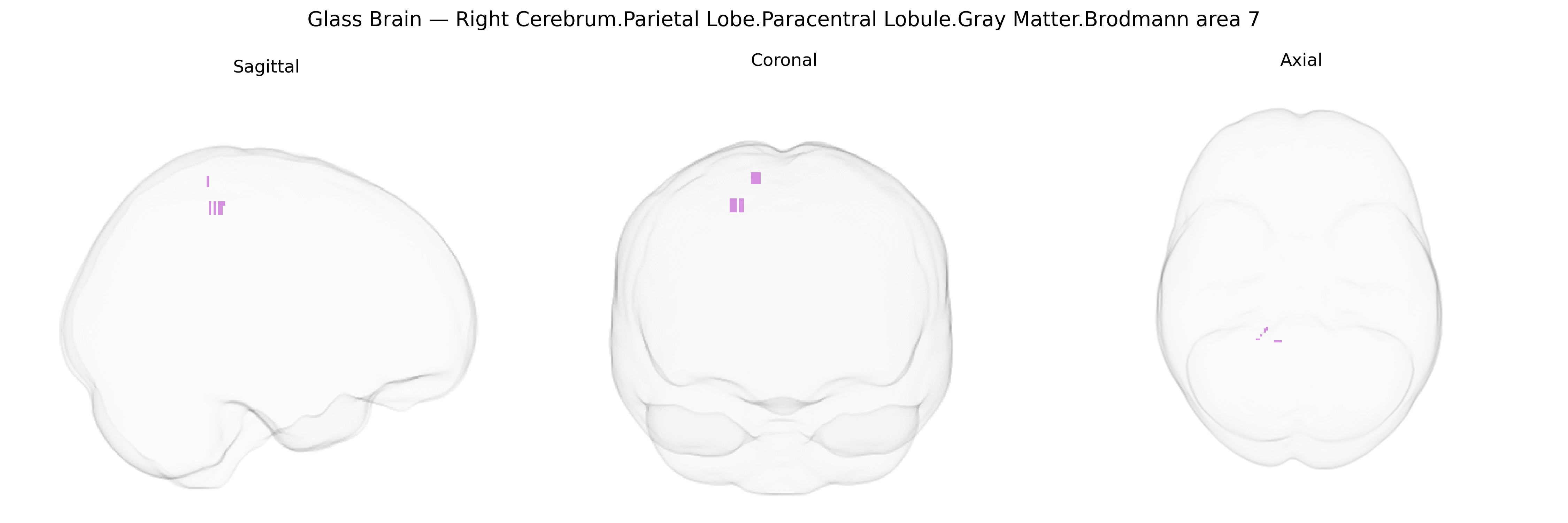
The bilateral Right Cerebrum.Parietal Lobe.Paracentral Lobule.Gray Matter.Brodmann area 7 corresponds to a superior parietal association cortical field situated near the medial extension of the superior parietal lobule, bordering regions involved in sensorimotor integration around the paracentral lobule. Brodmann area 7 is classically associated with higher-order somatosensory processing, visuospatial integration, and coordination of sensory information with motor planning, contributing to functions such as spatial attention, body schema, and guidance of visually directed movements. In the Talairach 1 mm Atlas, this label denotes gray matter in a multimodal association territory that integrates tactile, proprioceptive, and visual inputs to support goal-directed actions and complex spatial cognition. There is no direct link for this exact composite label; a related structure is Brodmann area 7.
The bilateral right paracentral lobule gray matter (including Brodmann area 7 in the superior parietal region as defined in the Talairach 1 mm atlas) has been implicated in multiple imaging genetics and GWAS-based brain mapping studies, though findings are typically reported at the level of parietal or sensorimotor cortices rather than this micro-anatomical label alone. Large-scale brain MRI GWAS consortia (e.g., ENIGMA, UK Biobank–based studies) have identified common variants in loci such as HMGA2, IGF1, microtubule and synaptic genes, and multiple neurodevelopmental pathways that influence total and regional cortical thickness and surface area in superior parietal and paracentral regions, including heritable variation in sensorimotor and somatosensory cortex morphology that overlaps this area. Genetic associations involving the paracentral lobule and adjacent superior parietal cortex have been reported in relation to motor and somatosensory traits, gait and balance measures, and individual differences in cognitive domains such as visuospatial attention and working memory, as well as in neurodevelopmental conditions (e.g., ADHD and autism spectrum disorder) and neurodegenerative diseases (especially Alzheimer’s disease and frontotemporal dementia), where risk variants in genes such as APOE, BIN1, and MAPT show downstream effects on parietal cortical atrophy patterns. Schizophrenia and major depression GWAS, via polygenic risk imaging analyses, have also linked higher polygenic load to structural and functional alterations in superior parietal and paracentral areas involved in sensorimotor integration and default-mode/somatomotor network coupling. However, most evidence is indirect and regionally coarse, and there is no single, well-established gene uniquely or specifically associated with the right paracentral lobule Brodmann area 7; rather, a broad polygenic architecture affecting cortical development, synaptic plasticity, and neurodegenerative pathways contributes to individual differences and disease-related changes in this region.
Overview generated by GPT-4o (2026).
Region ID: 1066
Hemisphere: bilateral
Atlas: Talairach labels 1mm
Full Quality Version: Download MP4
Full Quality Version: Download MP4
Wali Sidiqyar*, Gaurav Rudravaram*, Elyssa M. McMaster, Trent M. Schwartz, Adam M. Saunders, Kurt G. Schilling, Bennett A. Landman "Introducing SPINS: A Shared Public Visualization Library of Neuroanatomical Structures." Medical Imaging with Deep Learning- short paper
This resource is licensed under CC0 1.0 Universal (Public Domain).