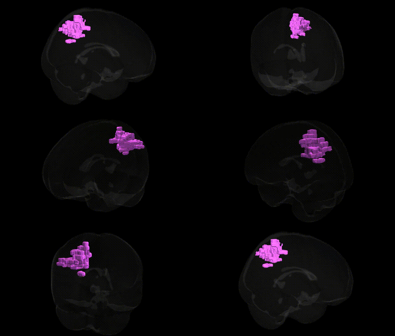
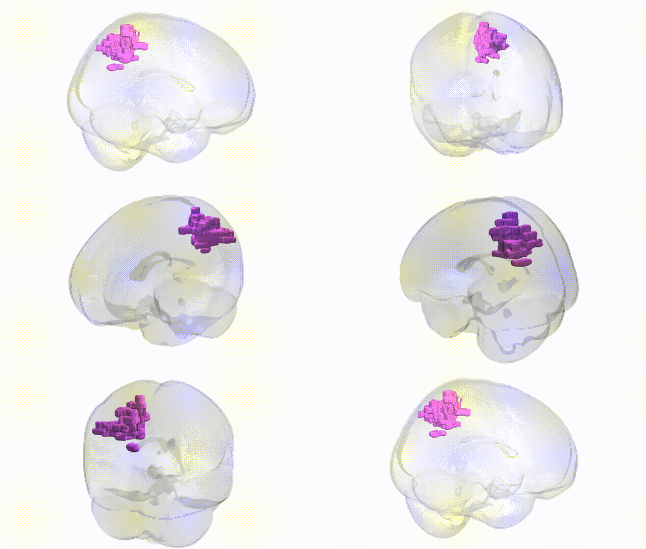
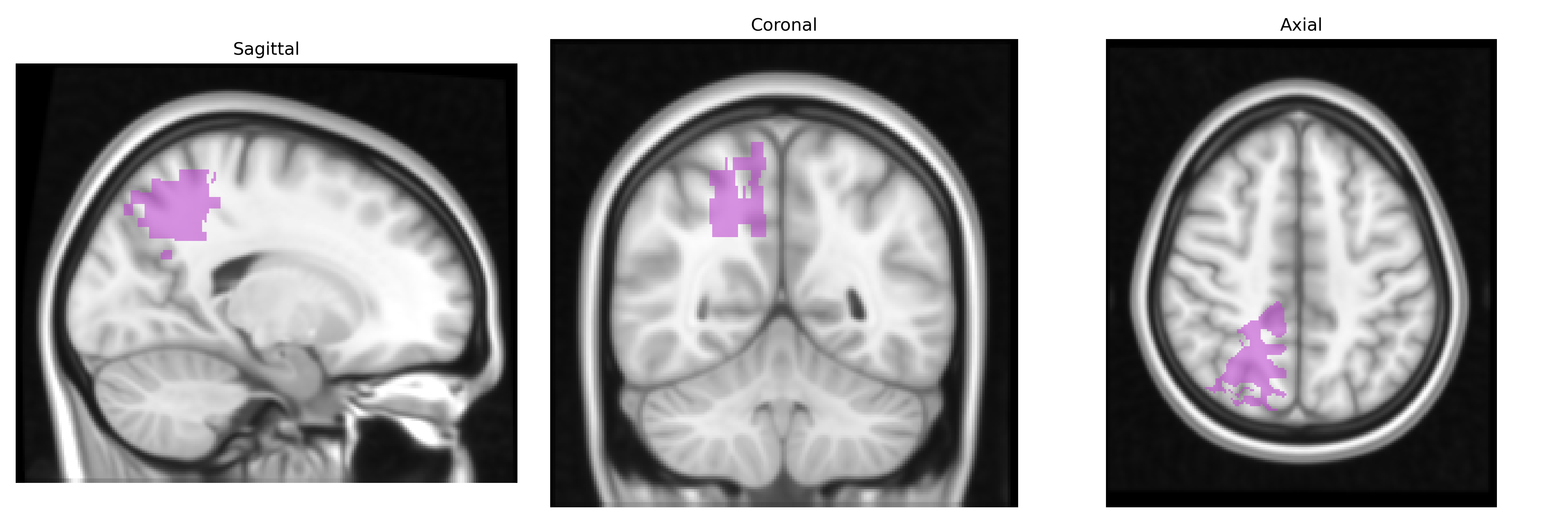
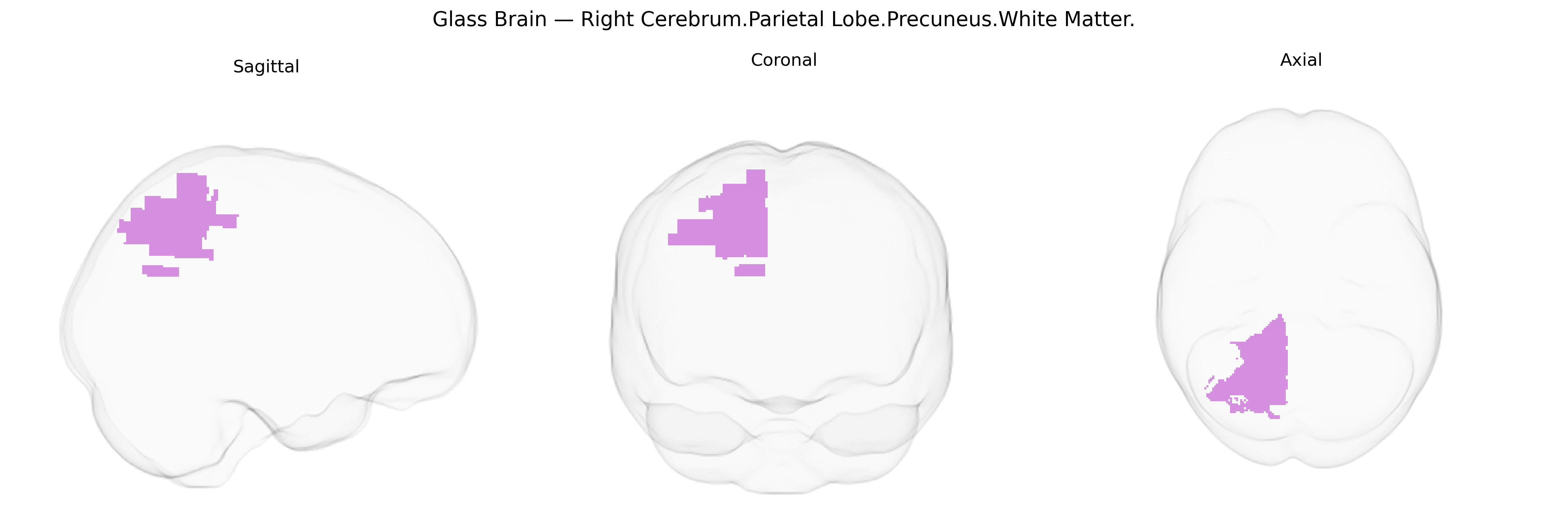
The bilateral Right Cerebrum.Parietal Lobe.Precuneus.White Matter corresponds to subcortical myelinated fiber pathways underlying the cortical precuneus within the medial parietal lobe of the right hemisphere, extending across both sides in the Talairach 1 mm labeling framework. These white matter tracts support communication between the precuneus cortex and distributed networks involving parietal, frontal, and occipital regions, contributing to higher-order integrative functions such as visuospatial processing, self-related cognition, and aspects of default mode network activity. The region lies along the medial wall of the parietal lobe, bordered by the cingulate sulcus and superior parietal lobule, and contains association fibers, callosal projections, and short U-fibers that integrate information within and between hemispheres. There is no direct Wikipedia article for the white matter of the precuneus; a related article describing the overlying cortical region is Precuneus.
The bilateral right precuneus white matter, as defined by the Talairach 1 mm atlas, participates in a broader parietal and default-mode network whose microstructure and connectivity show heritable variation and multiple genetic associations in imaging-genetics and GWAS studies. Twin and SNP-heritability work indicates substantial genetic influence on precuneus volume, cortical thickness, and white-matter integrity measures (e.g., fractional anisotropy) in adjacent tracts such as the superior longitudinal fasciculus and cingulum bundle, implicating genes involved in neurodevelopment, myelination, and synaptic function (including variants in genes like BDNF, NRG1, and others identified in large ENIGMA and UK Biobank analyses). GWAS of brain MRI phenotypes have reported associations between common variants and precuneus-related white-matter metrics, often overlapping with loci that also affect global intracranial volume and default-mode connectivity. Clinically, genetic risk for Alzheimer’s disease (notably APOE ε4 and polygenic risk scores) has been linked to altered metabolism, connectivity, and atrophy in the precuneus and its underlying white matter, and schizophrenia and major depressive disorder risk variants show convergent effects on precuneus structure and default-mode network function. Additional associations are reported for autism spectrum conditions, attention-deficit/hyperactivity disorder, and individual differences in cognitive traits such as episodic memory, self-referential processing, and mind-wandering, although many findings implicate the broader precuneus and parietal network rather than this precise Talairach-defined right white-matter label, and effect sizes are generally modest and polygenic.
Overview generated by GPT-4o (2026).
Region ID: 864
Hemisphere: bilateral
Atlas: Talairach labels 1mm
Full Quality Version: Download MP4
Full Quality Version: Download MP4
Wali Sidiqyar*, Gaurav Rudravaram*, Elyssa M. McMaster, Trent M. Schwartz, Adam M. Saunders, Kurt G. Schilling, Bennett A. Landman "Introducing SPINS: A Shared Public Visualization Library of Neuroanatomical Structures." Medical Imaging with Deep Learning- short paper
This resource is licensed under CC0 1.0 Universal (Public Domain).