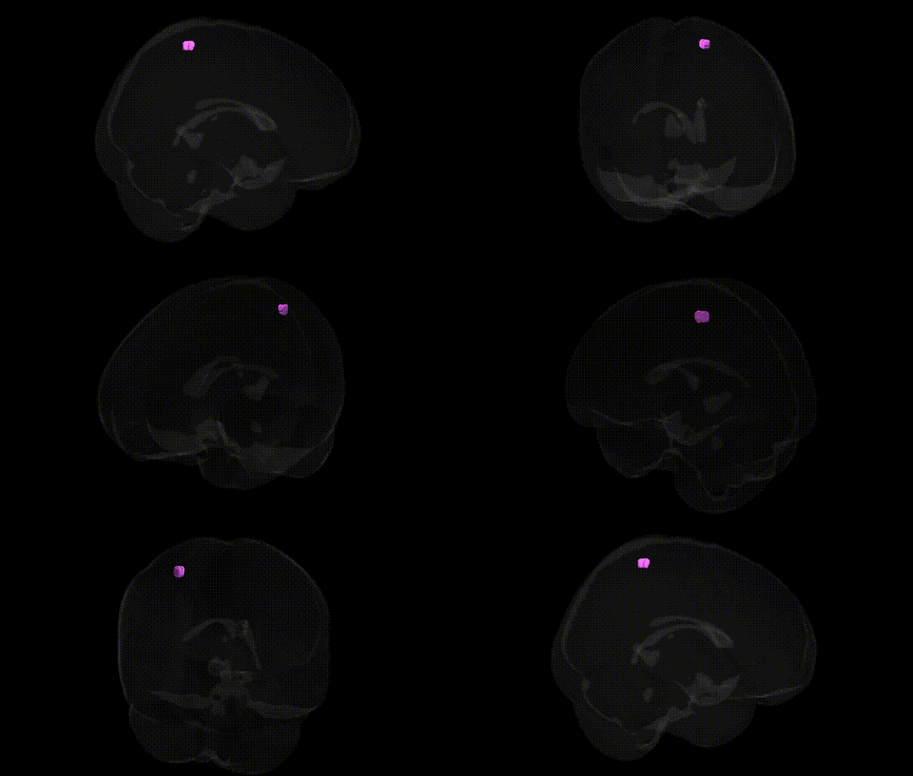
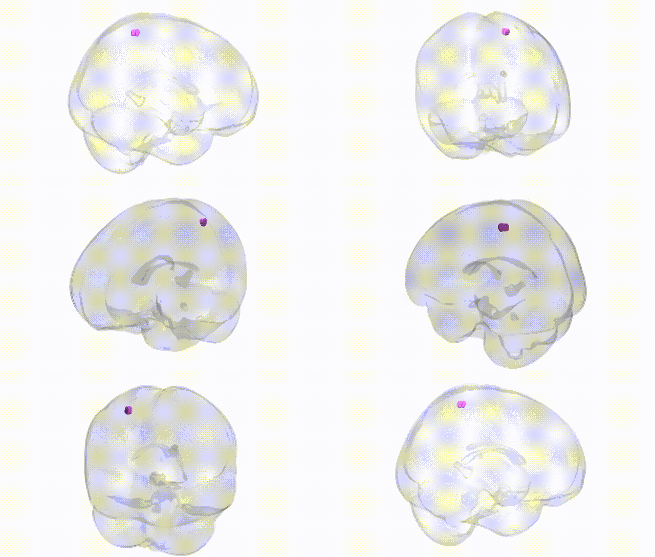
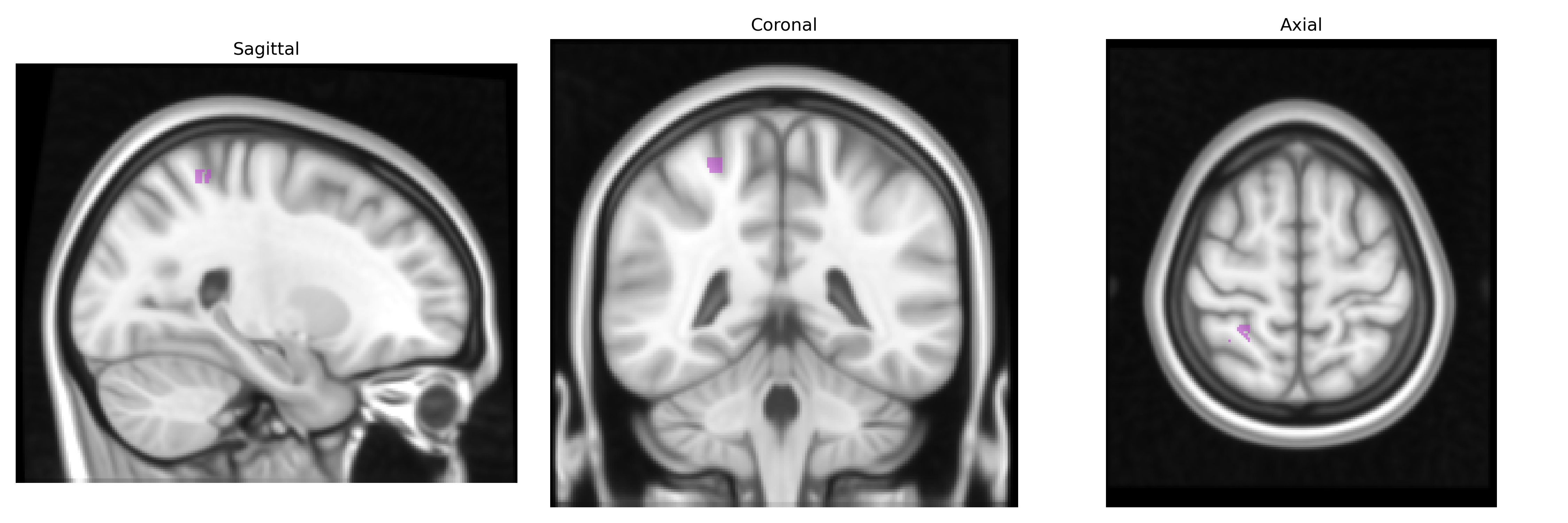
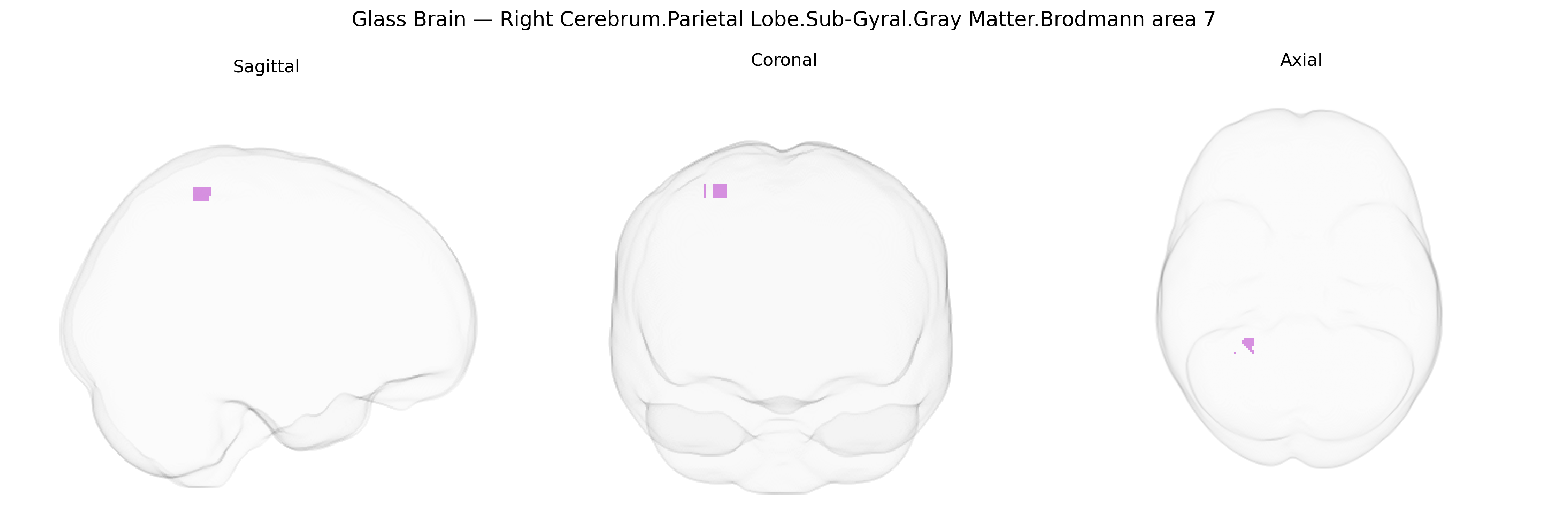
The bilateral Right Cerebrum.Parietal Lobe.Sub-Gyral.Gray Matter.Brodmann area 7 corresponds to a portion of the superior parietal lobule and adjacent precuneus in the dorsal posterior parietal cortex, characterized cytoarchitectonically by Brodmann area 7. This association cortex integrates multimodal sensory inputs, particularly visual and somatosensory information, to support visuospatial processing, spatial attention, sensorimotor integration, and the transformation of sensory coordinates into motor commands for actions such as reaching and grasping. Neurons in this region participate in constructing internal representations of extrapersonal space and body position, contributing to functions such as mental rotation, spatial working memory, and coordination of eye–hand movements. Lesions affecting this area can lead to deficits in spatial perception and attention, including elements of hemispatial neglect and impairments in complex visuomotor tasks. Brodmann area 7
The bilateral right parietal sub-gyral gray matter in Brodmann area 7, a core component of the superior parietal lobule implicated in visuospatial integration, attention, and sensorimotor coordination, has been indirectly linked to multiple genetic associations through imaging genetics and GWAS of brain structure and function. Large-scale neuroimaging GWAS (e.g., ENIGMA, UK Biobank) have identified variants in genes such as HMGA2, IGF1, MAPT, and multiple neurodevelopmental loci that influence parietal cortical thickness, surface area, and overall parietal volume, though these findings typically reference the superior parietal lobule rather than Brodmann area 7 at 1 mm Talairach resolution. Functional GWAS and polygenic score studies for cognitive traits (general intelligence, working memory, mathematical ability) frequently show parietal activation and structure–function correlations, with overlapping genetic signals in synaptic, neuroplasticity, and axon guidance pathways (e.g., genes involved in glutamatergic signaling and neuronal migration), suggesting shared genetic architecture between cognition and BA7 morphology. In neuropsychiatric and neurodevelopmental disorders, risk variants for schizophrenia, ADHD, and autism spectrum conditions—especially in genes affecting synaptic scaffolding (e.g., CACNA1C, GRIN2A, and other glutamate-related genes), immune signaling (e.g., complement pathway), and cortical development—have been associated with parietal abnormalities, including right-lateralized parietal thinning or altered connectivity involving BA7. Additionally, GWAS of Alzheimer’s disease and other dementias, including APOE and loci near BIN1 and CLU, have been linked to early structural and functional changes in posterior parietal regions, with BA7 often part of the affected posterior default-mode and dorsal attention networks. Although few studies report associations at the exact Talairach label “Right Cerebrum.Parietal Lobe.Sub-Gyral.Gray Matter.Brodmann area 7,” converging genetic evidence from structural and functional imaging GWAS indicates that variants influencing cortical development, synaptic signaling, and neurodegeneration contribute to inter-individual differences and disorder-related alterations in this parietal subregion.
Overview generated by GPT-4o (2026).
Region ID: 1081
Hemisphere: bilateral
Atlas: Talairach labels 1mm
Full Quality Version: Download MP4
Full Quality Version: Download MP4
Wali Sidiqyar*, Gaurav Rudravaram*, Elyssa M. McMaster, Trent M. Schwartz, Adam M. Saunders, Kurt G. Schilling, Bennett A. Landman "Introducing SPINS: A Shared Public Visualization Library of Neuroanatomical Structures." Medical Imaging with Deep Learning- short paper
This resource is licensed under CC0 1.0 Universal (Public Domain).