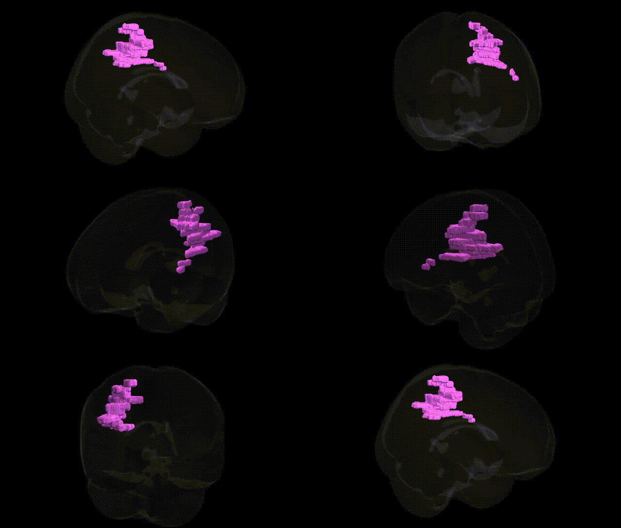
The bilateral Right Cerebrum.Parietal Lobe.Sub-Gyral.White Matter corresponds to deep parietal lobe white matter lying beneath the cortical gyri of the right parietal cortex, including portions underlying the superior and inferior parietal lobules and potentially the intraparietal sulcus. This region contains bundles of myelinated axons that form short association fibers linking neighboring parietal gyri, as well as longer association and projection pathways connecting parietal cortex with frontal, temporal, and occipital regions, and with subcortical structures via the internal capsule and related tracts. Functionally, these white matter pathways support multimodal sensory integration, visuospatial processing, attention, somatosensory discrimination, and aspects of sensorimotor coordination, with right-sided parietal connectivity particularly implicated in spatial awareness and attentional networks. There is no direct link for this exact sub-gyral white matter label; see the related structure Parietal lobe.
Genetic associations specifically targeting the bilateral Right Cerebrum.Parietal Lobe.Sub-Gyral.White Matter, as defined in the Talairach 1 mm atlas, are typically inferred from broader genome-wide association studies (GWAS) of parietal lobe structure, white-matter microstructure, and connectivity rather than from this exact label, with many findings coming from large consortia such as ENIGMA and UK Biobank imaging genetics. Variants in genes influencing axonal development, myelination, and synaptic function—such as those in or near CNTNAP2, NRG1, MAG, MBP, and other oligodendrocyte- and neuron-related loci—have been associated with parietal white-matter volume and diffusion metrics (e.g., fractional anisotropy) in tract-based studies that include sub-gyral parietal regions. GWAS of white-matter integrity and regional brain volumes have linked parietal white matter to polygenic risk for schizophrenia, major depressive disorder, bipolar disorder, and autism spectrum disorder, as well as to cognitive traits such as general intelligence, working memory, and educational attainment, with effects often mediated by widespread polygenic influences rather than single strong loci. In addition, risk variants for neurodegenerative and cerebrovascular disorders—such as APOE ε4 and other loci associated with Alzheimer’s disease and small-vessel disease—have been associated with reduced parietal white-matter integrity and posterior parietal disconnection patterns, implicating this region in the genetic architecture of dementia and age-related cognitive decline, though precise localization to the Talairach “sub-gyral” white matter label remains approximate rather than anatomically exclusive.
Overview generated by GPT-4o (2026).
Region ID: 798
Hemisphere: bilateral
Atlas: Talairach labels 1mm
Full Quality Version: Download MP4
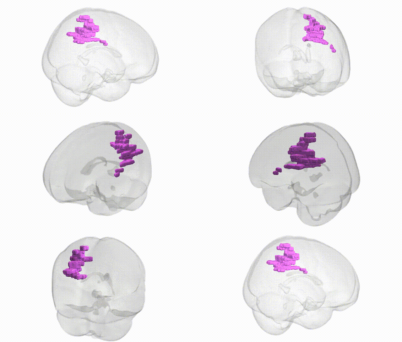
Full Quality Version: Download MP4
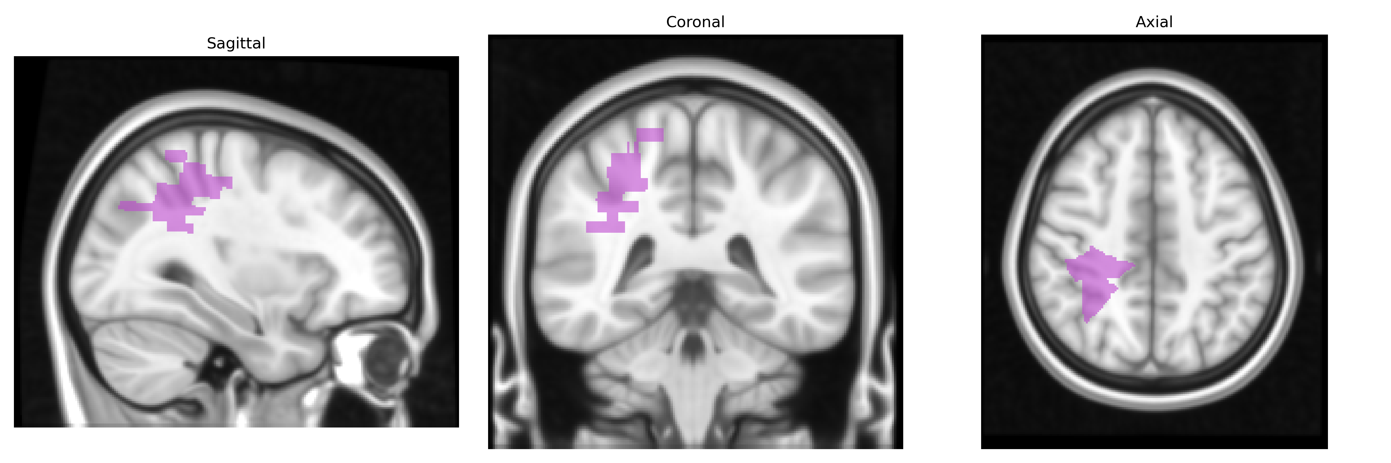
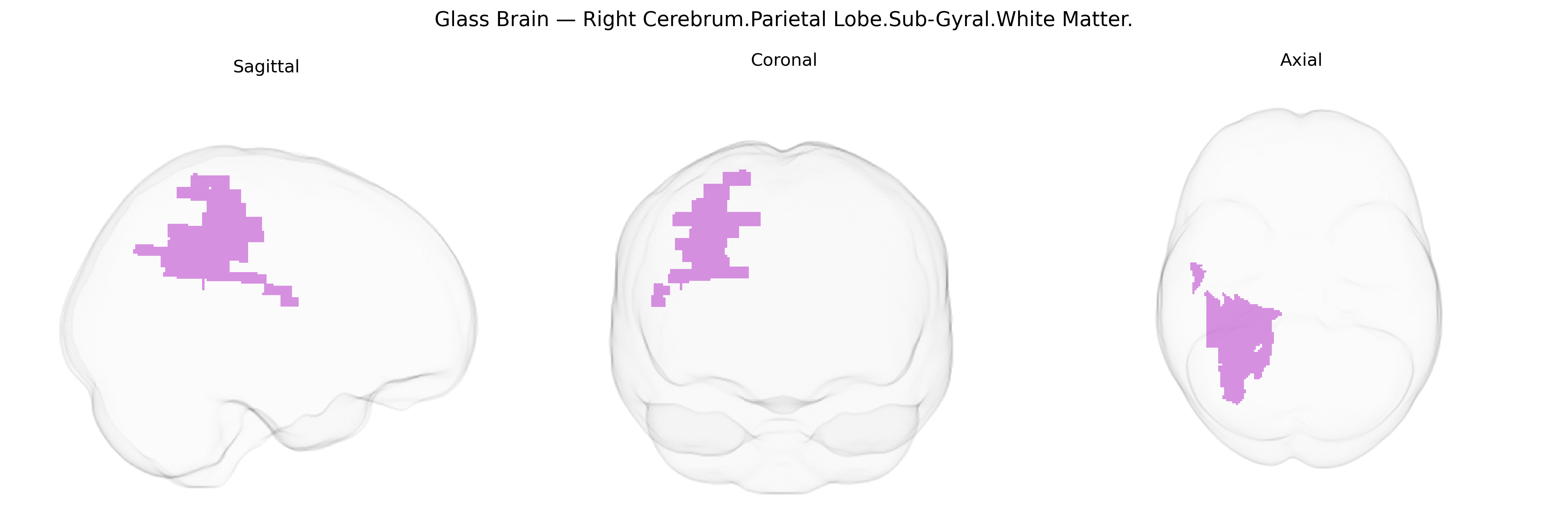
Wali Sidiqyar*, Gaurav Rudravaram*, Elyssa M. McMaster, Trent M. Schwartz, Adam M. Saunders, Kurt G. Schilling, Bennett A. Landman "Introducing SPINS: A Shared Public Visualization Library of Neuroanatomical Structures." Medical Imaging with Deep Learning- short paper
This resource is licensed under CC0 1.0 Universal (Public Domain).