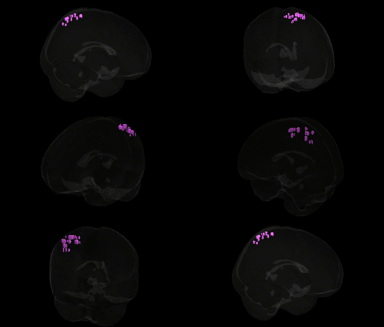
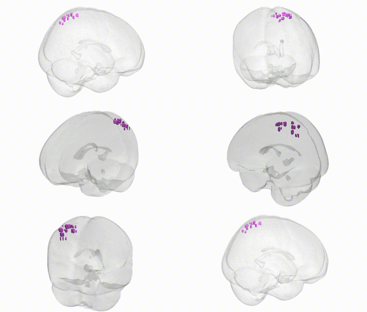
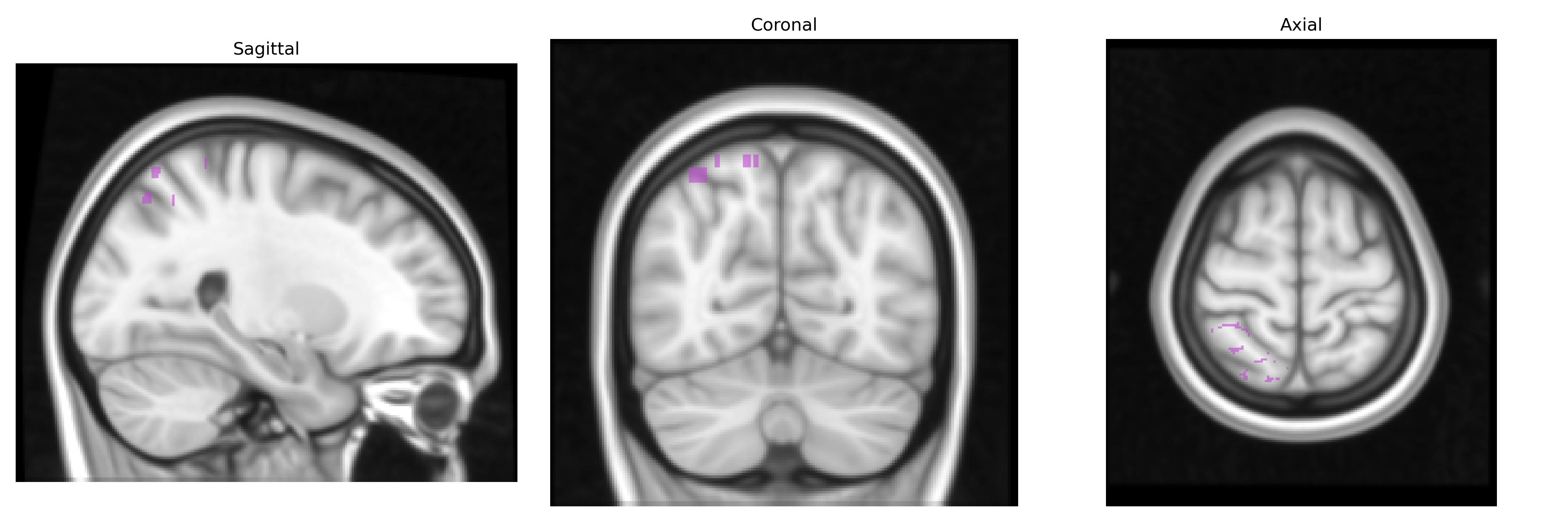
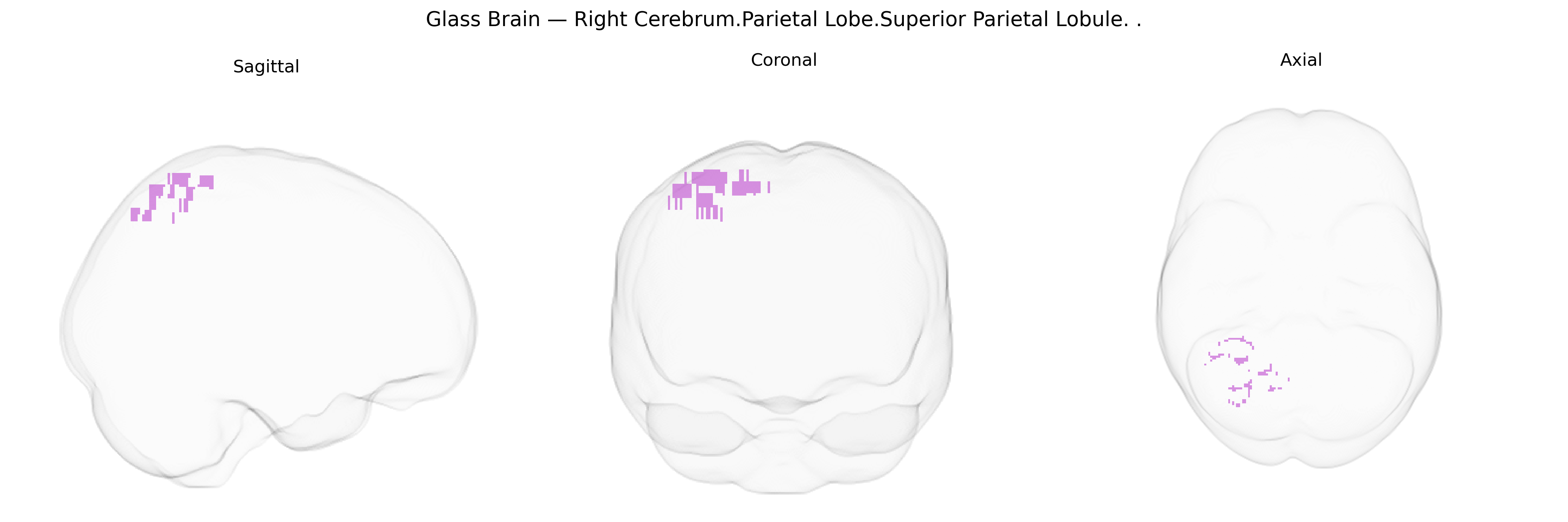
The bilateral Right Cerebrum.Parietal Lobe.Superior Parietal Lobule corresponds to the superior parietal lobule in the right hemisphere, a dorsal parietal association region implicated in multimodal sensory integration, spatial orientation, and attention. It receives convergent visual, somatosensory, and proprioceptive inputs and contributes to constructing body-centered and world-centered spatial representations, supporting functions such as visuomotor coordination, reaching, and object manipulation. This region is also involved in higher-order somatosensory processing, including discrimination of stimulus location and features, and interacts with frontal premotor and parietal networks involved in attention and working memory for spatial information. Superior parietal lobule
The bilateral right superior parietal lobule (SPL), as defined in the Talairach 1 mm atlas, has been implicated in multiple genetic and GWAS-based associations, primarily through large-scale imaging-genetics and neuropsychiatric studies that map variants to regional cortical thickness, surface area, or functional activation. Common variants in genes involved in synaptic plasticity, neurodevelopment, and axon guidance (for example, BDNF, NRG1, DISC1, and several calcium-channel and glutamatergic genes identified in ENIGMA and UK Biobank imaging GWAS) have been associated with structural variation in parietal regions encompassing the SPL, which in turn correlate with cognitive domains such as visuospatial attention, working memory, and general intelligence. GWAS of educational attainment and cognitive performance have repeatedly identified loci whose polygenic scores predict morphology and functional activation of the superior parietal cortex, suggesting a partially shared genetic architecture between parietal structure and cognitive ability. Parietal abnormalities involving the SPL are also reported in genetic studies of schizophrenia, bipolar disorder, major depressive disorder, autism spectrum disorder, and attention-deficit/hyperactivity disorder, where risk variants and polygenic risk scores often predict altered parietal volume or connectivity, although these effects are typically distributed and not specific to this region alone. Additionally, rare copy number variants and single-gene mutations linked to neurodevelopmental syndromes (for example, 22q11.2 deletion, and mutations affecting cortical patterning genes) can produce structural and functional alterations in the superior parietal cortex, supporting a broader genetic contribution to SPL development and function even though no single locus is uniquely or exclusively tied to the bilateral right superior parietal lobule.
Overview generated by GPT-4o (2026).
Region ID: 1038
Hemisphere: bilateral
Atlas: Talairach labels 1mm
Full Quality Version: Download MP4
Full Quality Version: Download MP4
Wali Sidiqyar*, Gaurav Rudravaram*, Elyssa M. McMaster, Trent M. Schwartz, Adam M. Saunders, Kurt G. Schilling, Bennett A. Landman "Introducing SPINS: A Shared Public Visualization Library of Neuroanatomical Structures." Medical Imaging with Deep Learning- short paper
This resource is licensed under CC0 1.0 Universal (Public Domain).