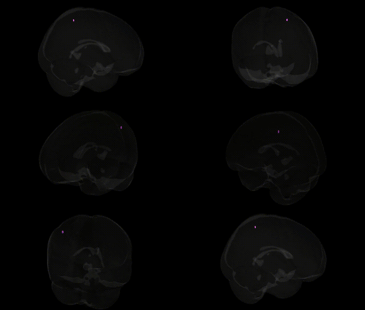
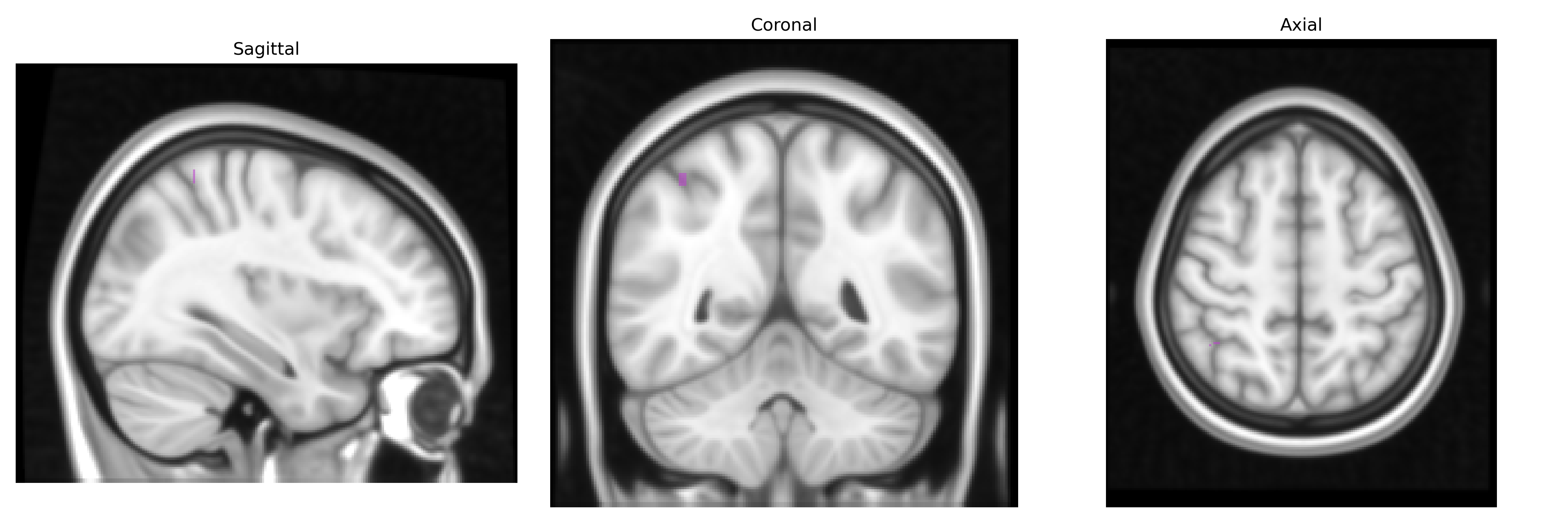
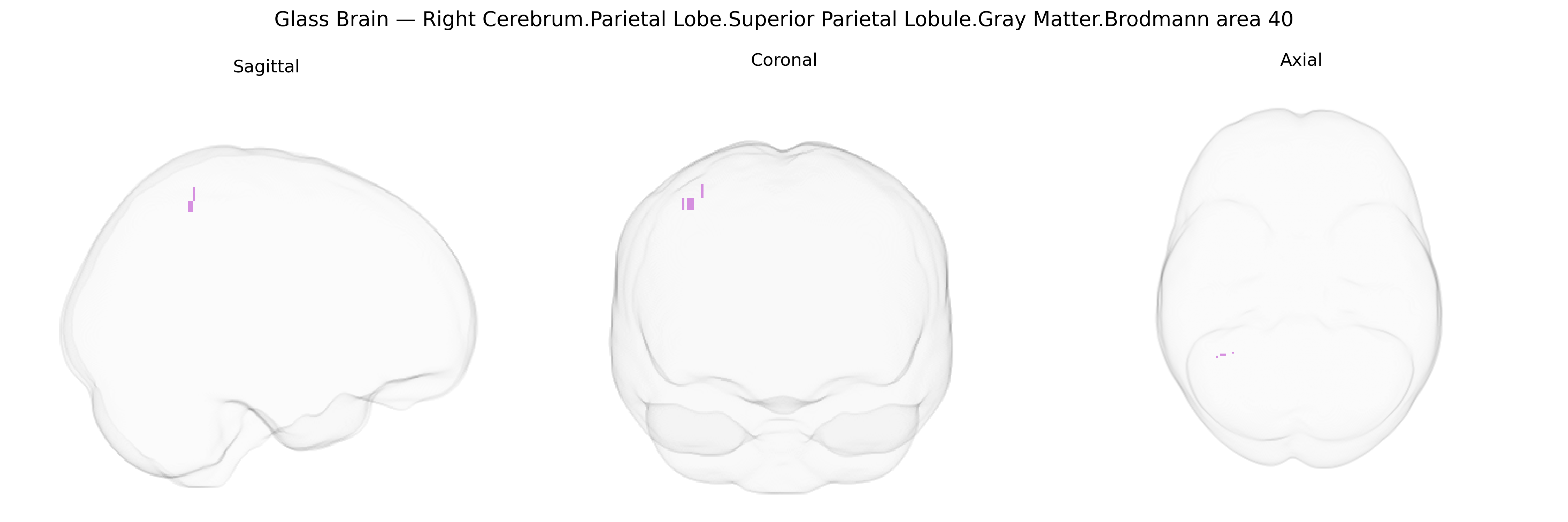
The bilateral Right Cerebrum Parietal Lobe Superior Parietal Lobule Gray Matter Brodmann area 40, as labeled in the Talairach 1 mm atlas, corresponds primarily to a portion of the inferior parietal cortex known as the supramarginal gyrus, a region traditionally associated with Brodmann area 40. This cortical field participates in multimodal integration of somatosensory, auditory, and visual information, contributing to functions such as language processing (especially phonological and reading-related tasks), spatial attention, praxis, and aspects of working memory. Neuronal populations in this area are involved in transforming sensory input into higher-order representations critical for object manipulation, body schema, and the coordination of perception and action, with strong connectivity to other parietal, temporal, and frontal association cortices.
The bilateral right superior parietal lobule (SPL) gray matter in or near Brodmann area 40, as defined by the Talairach 1 mm atlas, has been implicated in multiple genetic and GWAS-based associations through its roles in visuospatial attention, sensorimotor integration, working memory, and language-related processes; although BA40 is classically aligned with the supramarginal gyrus and inferior parietal lobule, many imaging-genetics and parcellation-based studies report overlapping or adjacent activation and structural variation in superior parietal regions. Polygenic and SNP-based studies have linked structural and functional variation in this right parietal territory to general cognitive ability and educational attainment (notably via large GWAS consortia such as ENIGMA and UK Biobank), with heritable differences in cortical thickness and surface area influenced by variants near genes involved in neurodevelopment and synaptic plasticity (e.g., MAPT region, microtubule and axon guidance genes). Imaging-genetics studies in schizophrenia, bipolar disorder, and major depressive disorder have repeatedly detected altered gray matter volume and connectivity in the right parietal cortex, with risk loci in genes related to glutamatergic signaling, calcium channels (e.g., CACNA1C and related loci), and immune-related pathways (e.g., MHC region) showing associations with parietal structural metrics or functional activation patterns. GWAS of ADHD and autism spectrum disorder have identified polygenic risk scores that predict atypical right parietal activation and morphology, particularly in tasks requiring attention shifting and social cognition, while Alzheimer’s disease and other dementias show heritable atrophy patterns that include right parietal regions, some of which co-localize with APOE and other neurodegeneration-related risk loci. Additionally, GWAS on traits such as handedness, visuospatial skills, and motor coordination have reported associations with variability in parietal cortex measures, suggesting that genetic influences on network-level organization of attention and sensorimotor systems partly converge on this right superior parietal/Brodmann area 40 territory, even though single-gene associations specific to this exact Talairach-defined parcel remain incompletely resolved and are typically inferred from broader parietal or frontoparietal network analyses rather than from highly localized region-of-interest GWAS.
Overview generated by GPT-4o (2026).
Region ID: 1062
Hemisphere: bilateral
Atlas: Talairach labels 1mm
Full Quality Version: Download MP4
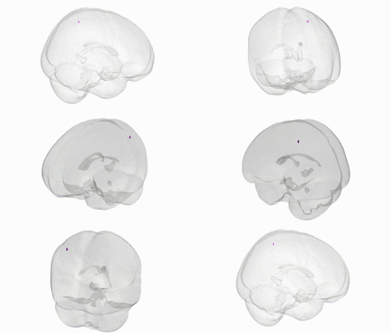
Full Quality Version: Download MP4
Wali Sidiqyar*, Gaurav Rudravaram*, Elyssa M. McMaster, Trent M. Schwartz, Adam M. Saunders, Kurt G. Schilling, Bennett A. Landman "Introducing SPINS: A Shared Public Visualization Library of Neuroanatomical Structures." Medical Imaging with Deep Learning- short paper
This resource is licensed under CC0 1.0 Universal (Public Domain).