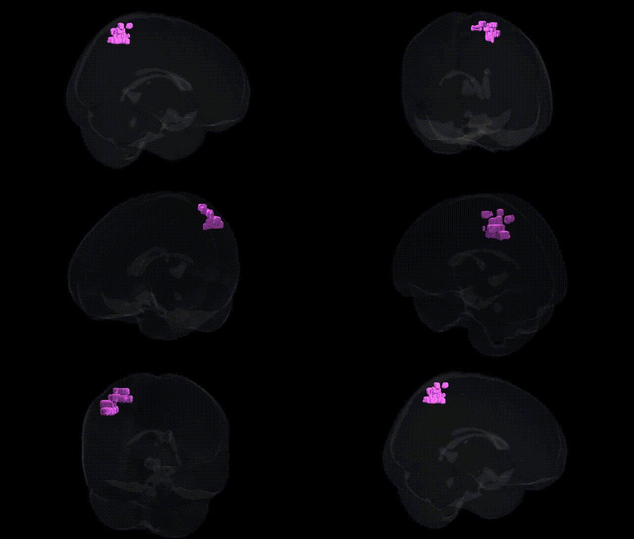
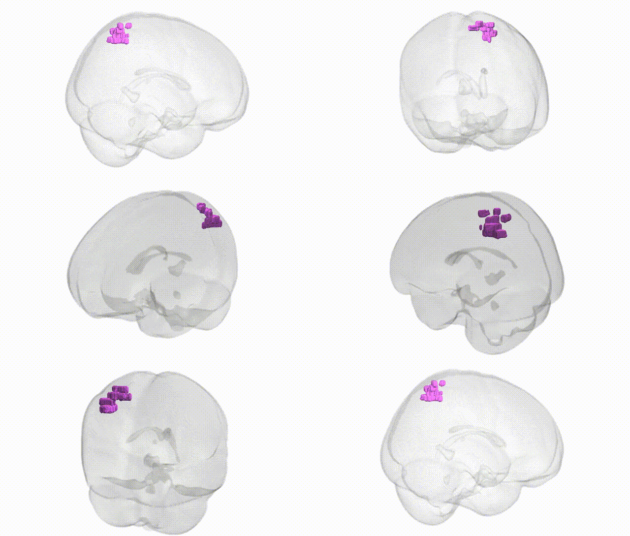
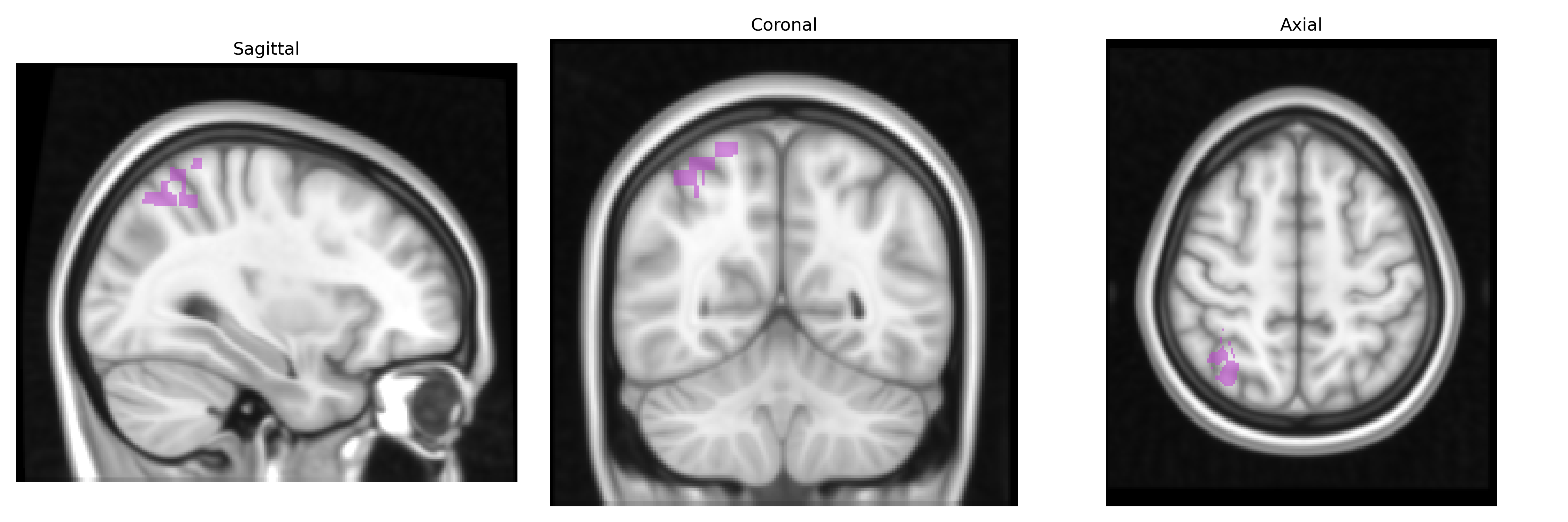
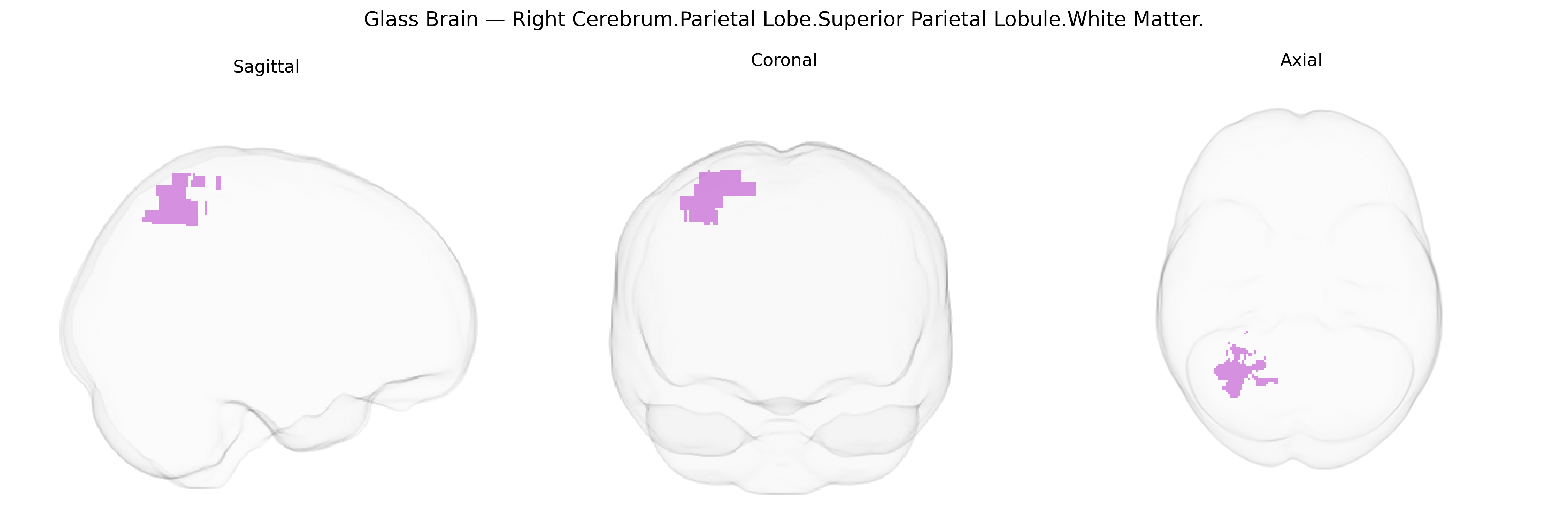
The bilateral Right Cerebrum.Parietal Lobe.Superior Parietal Lobule.White Matter comprises the deep myelinated fiber tracts underlying the superior parietal lobule in the right cerebral hemisphere, connecting it with neighboring parietal, frontal, occipital, and subcortical regions. This white matter supports integration of multimodal sensory information, visuospatial processing, and aspects of sensorimotor coordination by enabling efficient communication between cortical areas involved in spatial attention, body representation, and visually guided action. Tracts in this region contribute to long-range association pathways, including components of the superior longitudinal fasciculus, and are critical for the functional coupling of dorsal stream visuospatial networks. There is no direct link for this specific white matter subdivision; see the related cortical region: Superior parietal lobule.
The bilateral Right Cerebrum.Parietal Lobe.Superior Parietal Lobule.White Matter region, as defined in the Talairach labels 1 mm atlas, has been implicated indirectly in multiple genetic and GWAS findings through its involvement in large-scale white-matter tracts, particularly the superior longitudinal fasciculus and adjacent association fibers. Variants in genes affecting myelination and axonal integrity (e.g., MAG, MBP, PLP1, and other oligodendrocyte-related genes) have been associated with microstructural properties of superior parietal white matter in diffusion tensor imaging GWAS, including measures such as fractional anisotropy and mean diffusivity. Large neuroimaging-genetics consortia (e.g., ENIGMA, UK Biobank) have reported SNP-based heritability and specific loci influencing parietal white-matter volume and integrity, with signals often mapping to genes involved in neurodevelopment (e.g., genes within the 3p26, 17q21, and 22q11 regions) and axon guidance. Disorders in which GWAS or imaging–genetic studies highlight superior parietal white matter include schizophrenia and bipolar disorder (with risk loci in synaptic and neurodevelopmental genes showing associated parietal white-matter abnormalities), autism spectrum disorder (where rare CNVs and polygenic burden correlate with altered parietal connectivity), attention-deficit/hyperactivity disorder and general cognitive ability (g) (where polygenic scores for education and cognition correlate with superior parietal white-matter structure), and neurodegenerative diseases such as Alzheimer’s disease, in which APOE ε4 and other risk variants relate to early white-matter changes in posterior parietal networks. Overall, genetic associations for this specific atlas-defined region are typically inferred from tract-based or lobar analyses rather than region-exclusive GWAS, but consistently converge on genes and polygenic profiles governing neurodevelopment, myelination, and higher-order cognitive and visuospatial networks in which the superior parietal white matter plays a central role.
Overview generated by GPT-4o (2026).
Region ID: 1042
Hemisphere: bilateral
Atlas: Talairach labels 1mm
Full Quality Version: Download MP4
Full Quality Version: Download MP4
Wali Sidiqyar*, Gaurav Rudravaram*, Elyssa M. McMaster, Trent M. Schwartz, Adam M. Saunders, Kurt G. Schilling, Bennett A. Landman "Introducing SPINS: A Shared Public Visualization Library of Neuroanatomical Structures." Medical Imaging with Deep Learning- short paper
This resource is licensed under CC0 1.0 Universal (Public Domain).