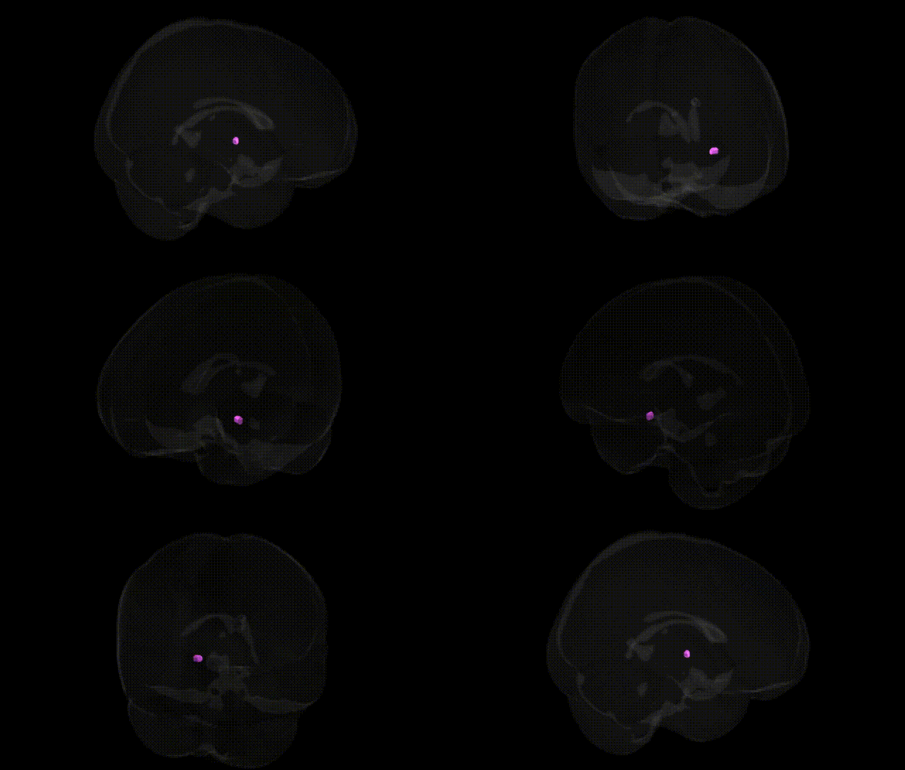
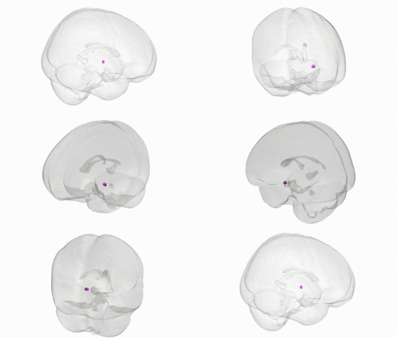
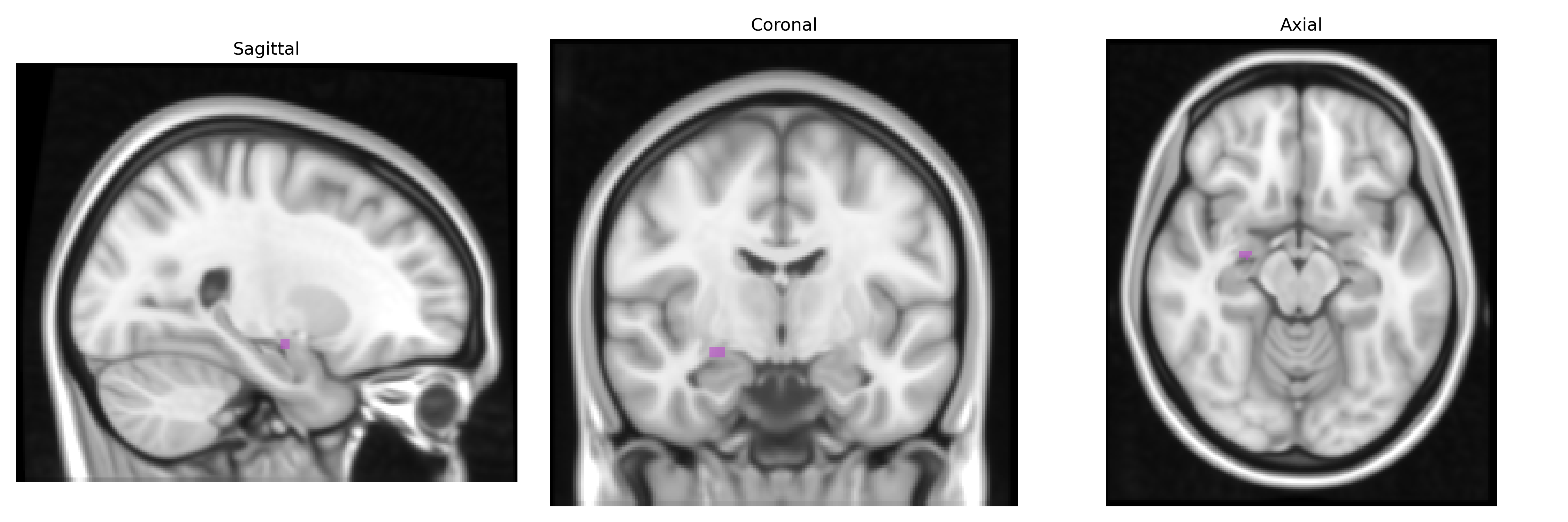
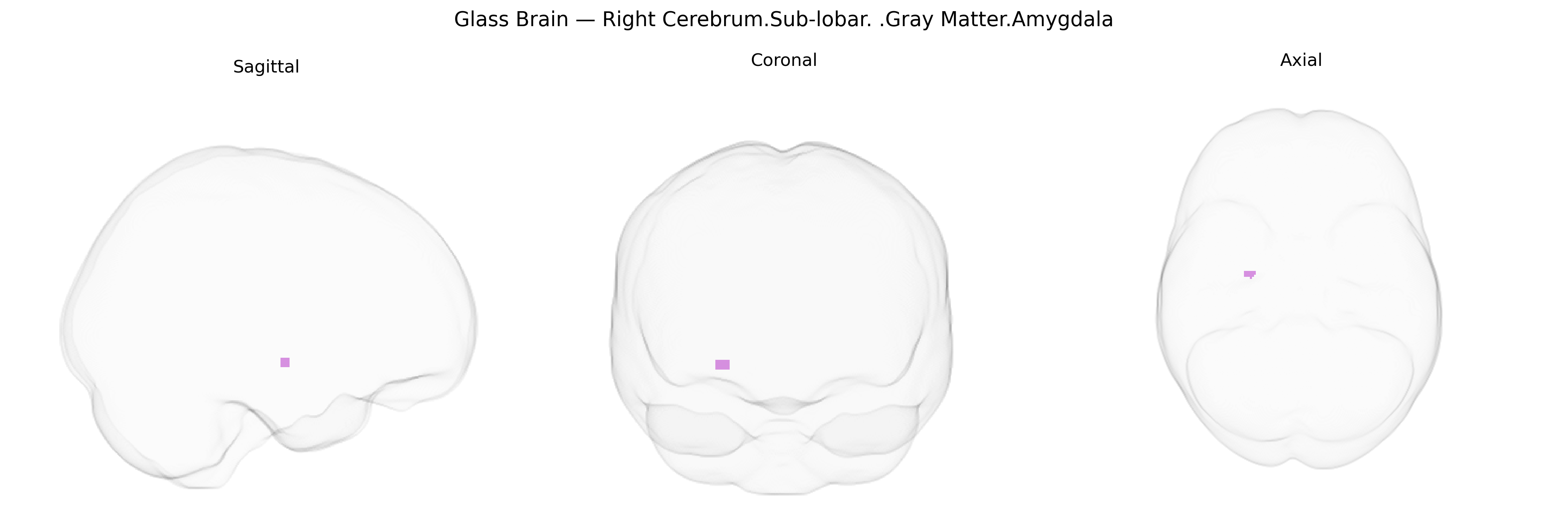
The bilateral Right Cerebrum.Sub-lobar.Gray Matter.Amygdala, as defined in the Talairach 1 mm Atlas, corresponds to the right amygdaloid complex, a collection of nuclei located in the medial temporal lobe deep to the uncus and anterior to the hippocampus. This gray-matter structure is a central hub for processing emotional salience, especially fear, threat detection, and reward-related cues, and contributes critically to emotional learning, associative memory, and modulation of autonomic and endocrine responses via its connections with the hypothalamus and brainstem. Extensive reciprocal connections with the prefrontal cortex, hippocampus, sensory association cortices, and other limbic structures enable integration of sensory input with past experience to guide behavioral and visceral responses; lateralization to the right hemisphere has been associated in some studies with preferential roles in rapid, automatic emotional evaluation and negative affect. Amygdala
The bilateral right sub-lobar amygdala (Talairach 1 mm atlas) shows robust genetic associations from imaging–genetics and GWAS studies implicating it in psychiatric, neurodevelopmental, and neurodegenerative phenotypes: common variants in genes related to serotonin (e.g., SLC6A4/5-HTTLPR), dopamine (e.g., DRD2, COMT), neurotrophins (e.g., BDNF Val66Met), and stress-response systems (e.g., FKBP5, CRHR1) have been linked to individual differences in amygdala gray-matter volume and reactivity, often with right-lateralized or bilateral effects; large-scale imaging GWAS (e.g., ENIGMA, UK Biobank) have identified SNPs across multiple loci (including genes involved in synaptic function, cell adhesion, and neurodevelopment) that influence amygdala volume and shape, though specific hits vary by study and threshold. Genetically modulated amygdala structure and function is associated with risk for major depressive disorder, generalized and social anxiety disorders, post-traumatic stress disorder, bipolar disorder, and schizophrenia, as well as traits such as neuroticism, extraversion, harm avoidance, and sensitivity to emotional stimuli, with polygenic risk scores for depression, schizophrenia, and autism spectrum conditions showing correlations with amygdala morphology and activation; additional associations extend to Alzheimer’s disease and other dementias, where risk variants (e.g., APOE ε4) are linked to amygdala atrophy and altered connectivity. Overall, the right amygdala emerges as a heritable, genetically modulated hub in circuits supporting fear, affective salience, and social-emotional processing, with convergent evidence from GWAS and candidate-gene work tying its structure and function to genetic liability for multiple brain-based disorders and personality traits.
Overview generated by GPT-4o (2026).
Region ID: 358
Hemisphere: bilateral
Atlas: Talairach labels 1mm
Full Quality Version: Download MP4
Full Quality Version: Download MP4
Wali Sidiqyar*, Gaurav Rudravaram*, Elyssa M. McMaster, Trent M. Schwartz, Adam M. Saunders, Kurt G. Schilling, Bennett A. Landman "Introducing SPINS: A Shared Public Visualization Library of Neuroanatomical Structures." Medical Imaging with Deep Learning- short paper
This resource is licensed under CC0 1.0 Universal (Public Domain).