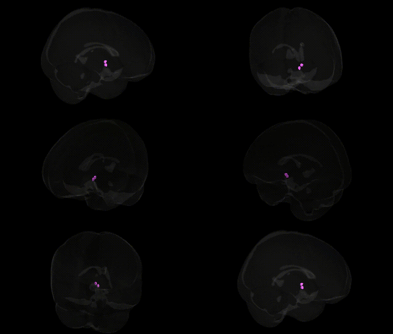
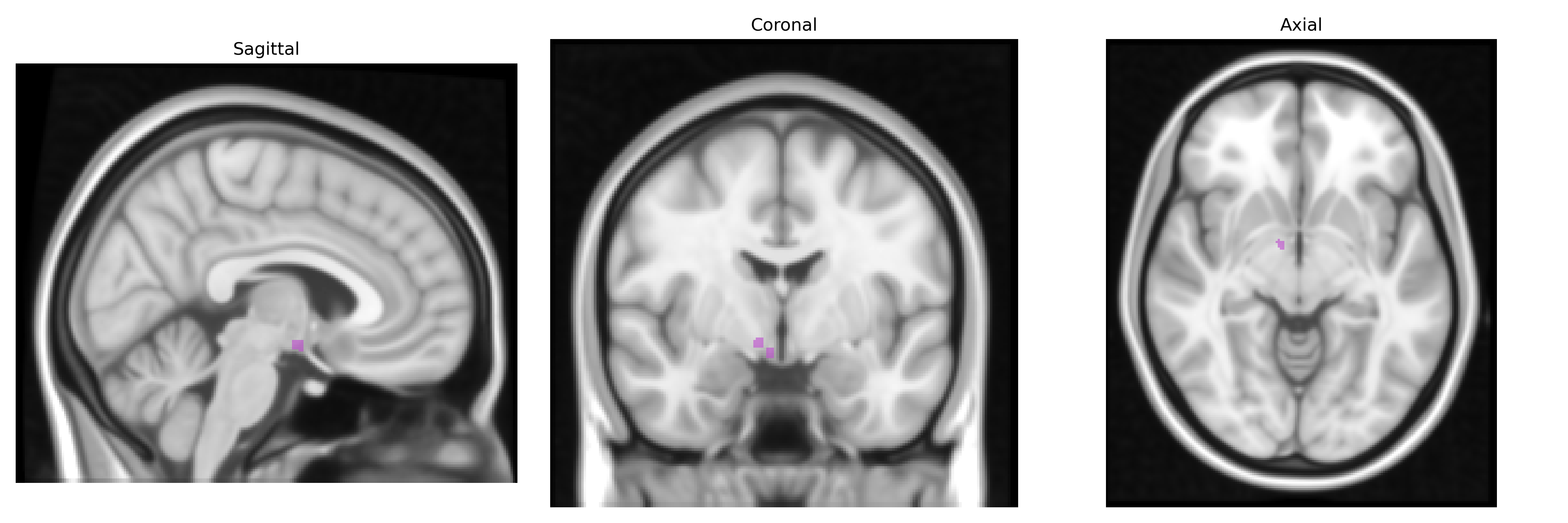
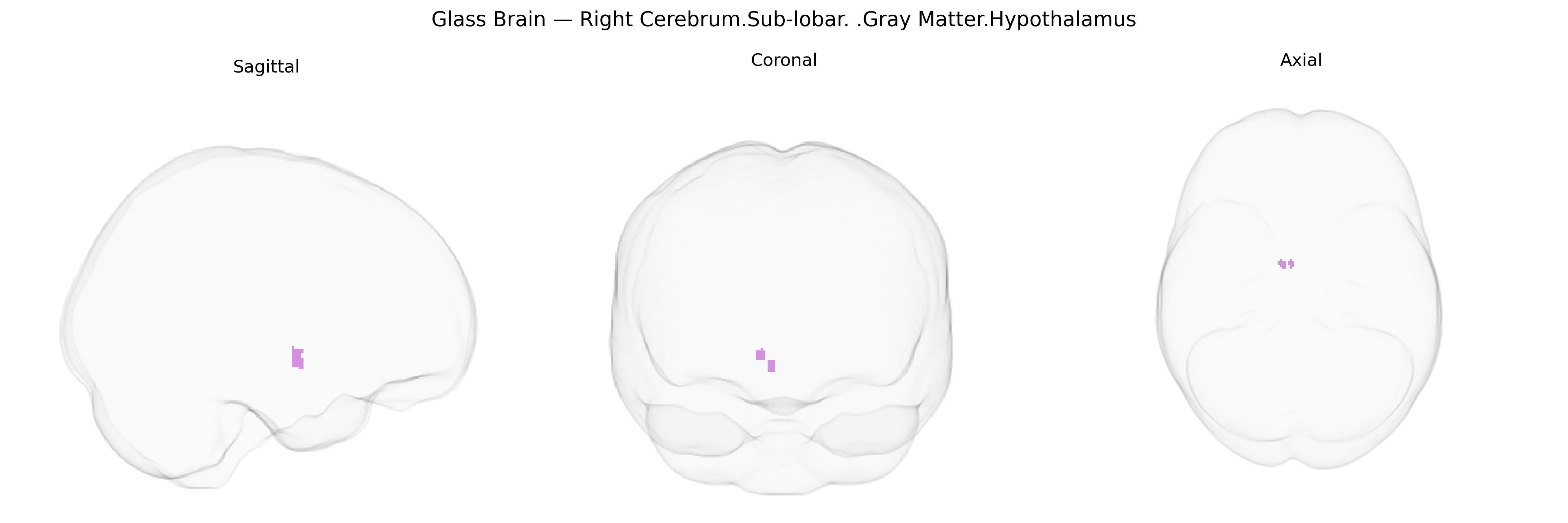
The bilateral Right Cerebrum.Sub-lobar.Gray Matter.Hypothalamus region in the Talairach 1 mm atlas corresponds to hypothalamic gray matter located within the sub-lobar territory of the right cerebral hemisphere and mirrored on the left, forming a midline, diencephalic structure at the base of the brain, below the thalamus and bordering the third ventricle. This region comprises multiple nuclei and fiber pathways that integrate autonomic, endocrine, and behavioral responses critical for homeostasis, including regulation of body temperature, circadian rhythms, feeding and energy balance, stress responses, fluid balance, and reproductive and defensive behaviors. It interfaces with limbic, brainstem, and cortical circuits, and exerts major influence on the pituitary gland via neuroendocrine projections, thereby coordinating hormonal outputs with neural activity. Hypothalamus
The hypothalamus, as defined in the Talairach 1 mm atlas within the Right Cerebrum.Sub-lobar.Gray Matter region (bilateral), has been implicated in multiple genetic association studies spanning neuropsychiatric, metabolic, and neurodegenerative traits, although few GWAS directly target this specific atlas label; instead, most findings come from imaging-genetics and region-of-interest analyses that include hypothalamic volume or function. Polygenic variation in genes regulating energy homeostasis and neuroendocrine signaling—such as MC4R, LEPR, BDNF, FTO, and genes in the melanocortin and insulin signaling pathways—has been repeatedly associated with hypothalamic structure or activity and with obesity, type 2 diabetes, and related metabolic phenotypes. Large-scale imaging GWAS (e.g., ENIGMA and UK Biobank–based studies) report SNP heritability for hypothalamic volume and identify loci near or within developmental and axon-guidance genes, as well as genes involved in neuronal differentiation, although the specific lead variants and mapped genes vary across studies. Hypothalamic alterations linked to genetic risk scores have been reported in major depressive disorder, anxiety, bipolar disorder, schizophrenia, and PTSD, particularly in relation to stress-response and HPA-axis genes (e.g., CRHR1, FKBP5, NR3C1), and in neurodegenerative disorders such as Alzheimer’s disease, where APOE and other AD risk loci correlate with hypothalamic atrophy or metabolic dysregulation. Additional associations involve circadian rhythm and sleep-related genes (e.g., CLOCK, PER3, ARNTL) affecting hypothalamus-mediated sleep–wake and thermoregulatory functions, as well as reproductive and pubertal timing loci (e.g., KISS1, GNRH1 pathway genes) influencing hypothalamic–pituitary–gonadal axis regulation. Overall, genetic studies support a highly polygenic architecture for hypothalamic structure and function, with pleiotropic loci that jointly affect metabolic, endocrine, affective, and cognitive phenotypes through this sub-lobar gray matter region.
Overview generated by GPT-4o (2026).
Region ID: 364
Hemisphere: bilateral
Atlas: Talairach labels 1mm
Full Quality Version: Download MP4
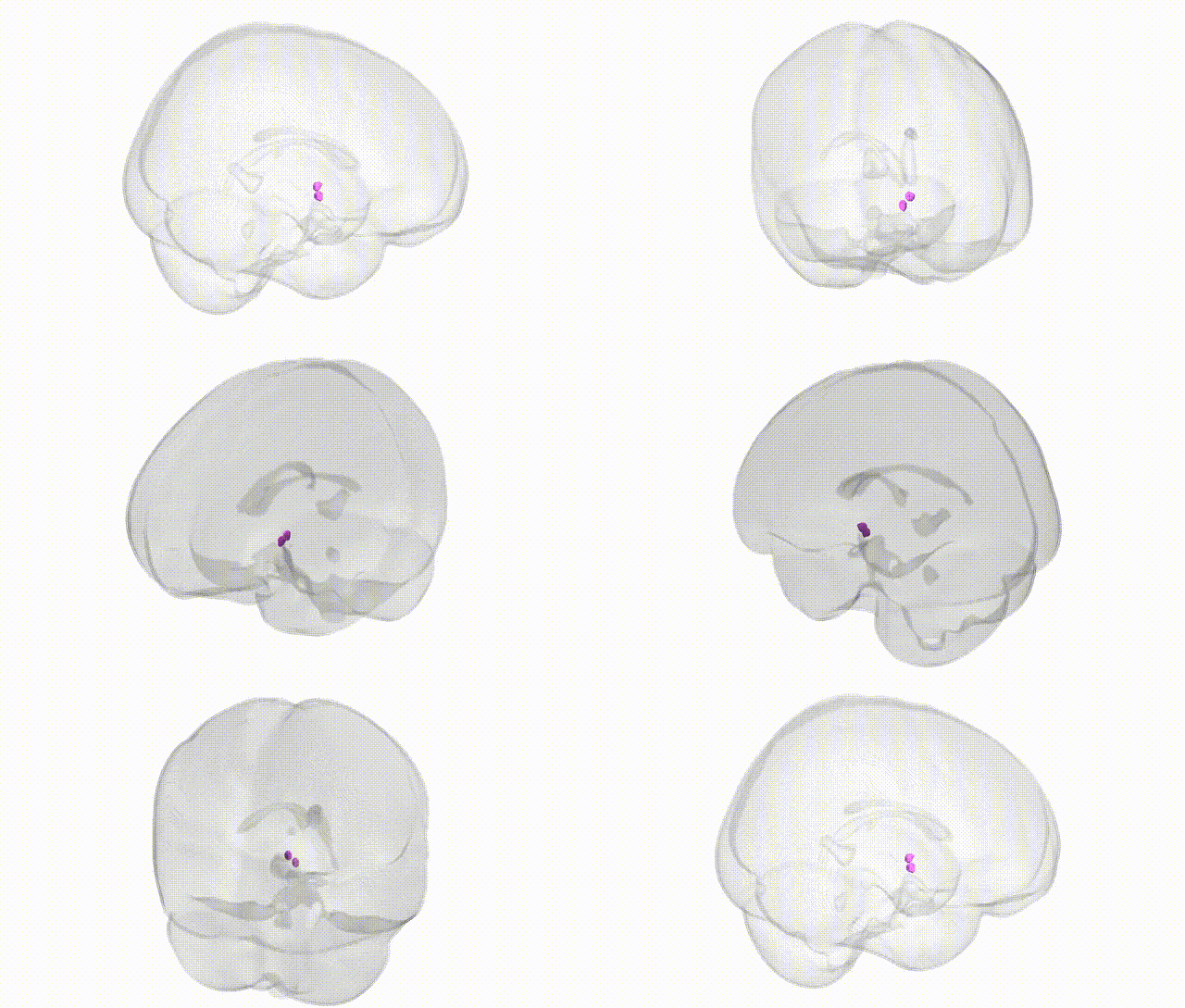
Full Quality Version: Download MP4
Wali Sidiqyar*, Gaurav Rudravaram*, Elyssa M. McMaster, Trent M. Schwartz, Adam M. Saunders, Kurt G. Schilling, Bennett A. Landman "Introducing SPINS: A Shared Public Visualization Library of Neuroanatomical Structures." Medical Imaging with Deep Learning- short paper
This resource is licensed under CC0 1.0 Universal (Public Domain).