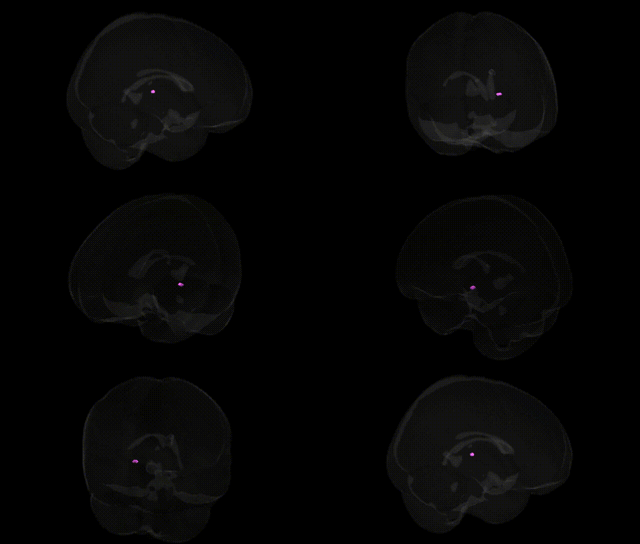
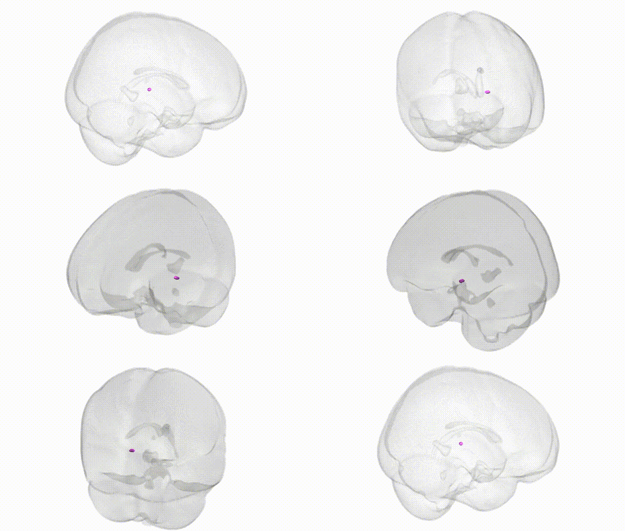
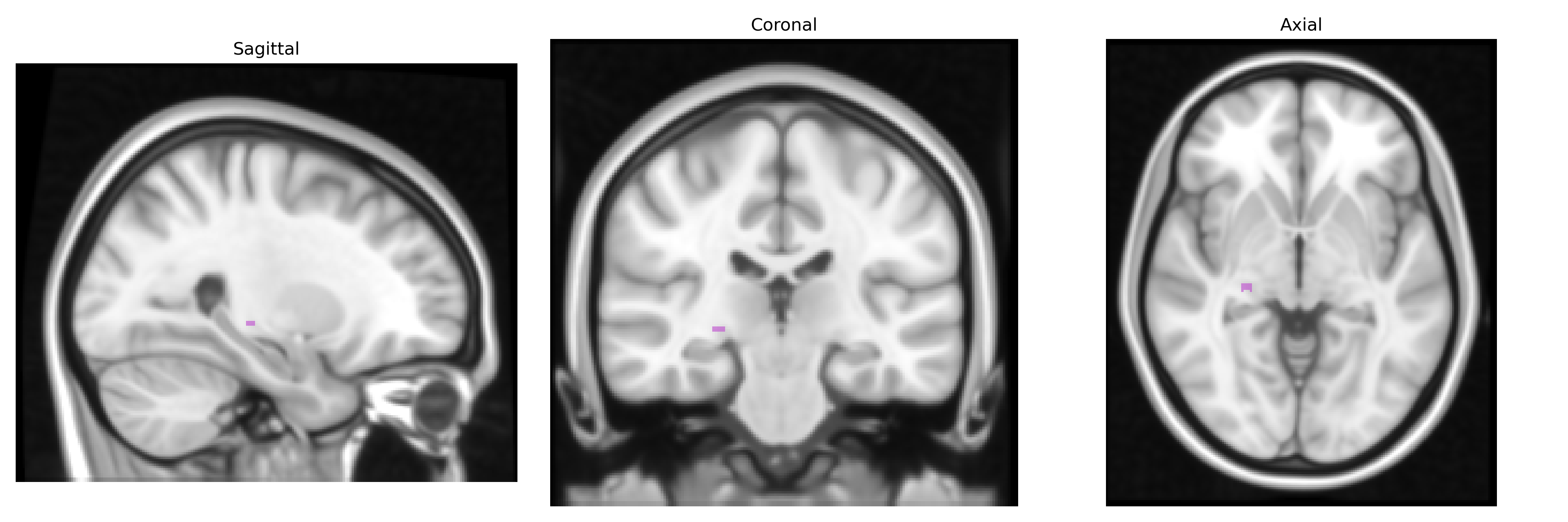
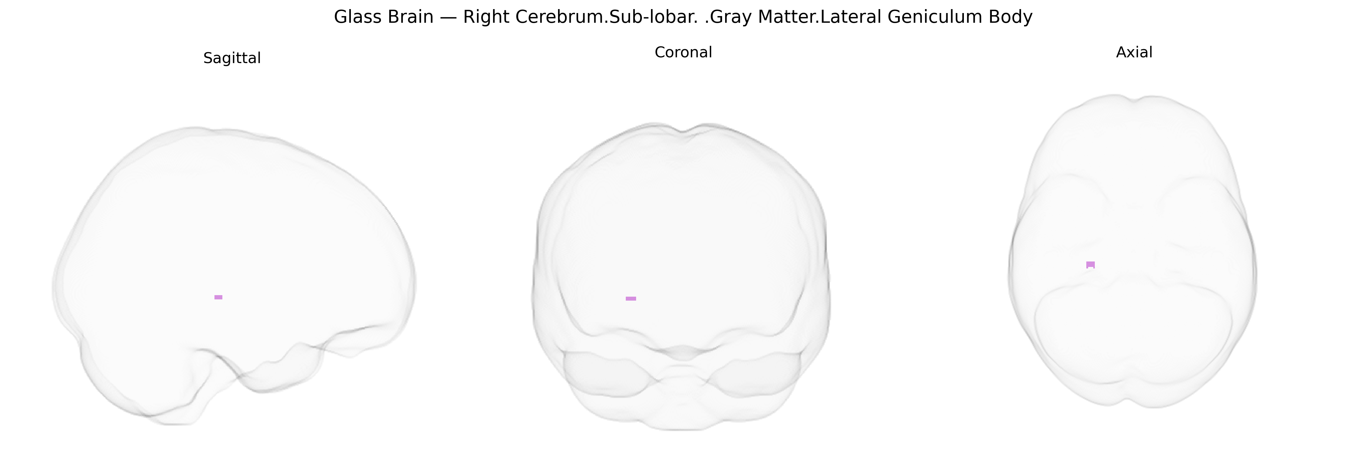
The bilateral Right Cerebrum.Sub-lobar.Gray Matter.Lateral Geniculum Body corresponds to the lateral geniculate nucleus (LGN) of the thalamus, a laminated subcortical relay center in the visual pathway located in the posterolateral thalamus beneath the cortical surface. It receives segregated, topographically organized retinal input via the optic tract and projects to primary visual cortex (V1, Brodmann area 17) through the optic radiation, preserving information about eye of origin, spatial location, and distinct visual channels (magnocellular and parvocellular pathways) important for motion, depth, color, and fine detail processing. As part of the sub-lobar gray matter, it participates in early processing and gating of visual information, contributes to binocular vision and visual attention, and integrates modulatory feedback from cortex and other structures, thereby influencing how visual signals are filtered and transmitted to cortical areas for conscious perception. Lateral geniculate nucleus
The lateral geniculate body (lateral geniculum), a thalamic relay nucleus for visual information, has been implicated in several genetic and imaging-genetics studies, although often under broader thalamic or subcortical volumetric measures rather than this specific Talairach label. GWAS of subcortical brain volumes from large consortia such as ENIGMA and UK Biobank have identified loci near genes involved in neurodevelopment, synaptic function, and axon guidance (for example, variants near NPTX1, NKAIN2, and other neurodevelopmental genes) associated with thalamic or thalamic subregion size, which would encompass the lateral geniculate complex. Structural and functional alterations of the lateral geniculate nucleus have been described in disorders with known genetic risk architectures, including multiple sclerosis (e.g., HLA-DRB1*15:01 and other immune-related loci), glaucoma and optic neuropathies (MYOC, OPTN, and mitochondrial genes in Leber hereditary optic neuropathy), and neurodevelopmental or psychiatric disorders such as schizophrenia and autism spectrum disorder, where polygenic risk scores and specific variants in genes governing synaptic plasticity and neuronal connectivity (e.g., CACNA1C, GRIN2A, and other glutamatergic and calcium-channel genes) show associations with thalamic and visual pathway structure or function. Visual system–focused GWAS, including those on visual acuity, refractive error, and amblyopia, implicate genes regulating retinal development and axonal projection (e.g., ATOH7, GJD2, and other optic nerve pathway genes), which, although acting primarily at the retinal and optic nerve levels, have downstream structural and functional correlates in the lateral geniculate nucleus. Overall, current genetic evidence links variation in neurodevelopmental, immune, and visual pathway genes to structural or functional changes in thalamic and lateral geniculate regions, typically via broader imaging-genetics analyses rather than region-specific GWAS limited solely to the bilateral Right Cerebrum.Sub-lobar.Gray Matter.Lateral Geniculum Body label.
Overview generated by GPT-4o (2026).
Region ID: 500
Hemisphere: bilateral
Atlas: Talairach labels 1mm
Full Quality Version: Download MP4
Full Quality Version: Download MP4
Wali Sidiqyar*, Gaurav Rudravaram*, Elyssa M. McMaster, Trent M. Schwartz, Adam M. Saunders, Kurt G. Schilling, Bennett A. Landman "Introducing SPINS: A Shared Public Visualization Library of Neuroanatomical Structures." Medical Imaging with Deep Learning- short paper
This resource is licensed under CC0 1.0 Universal (Public Domain).