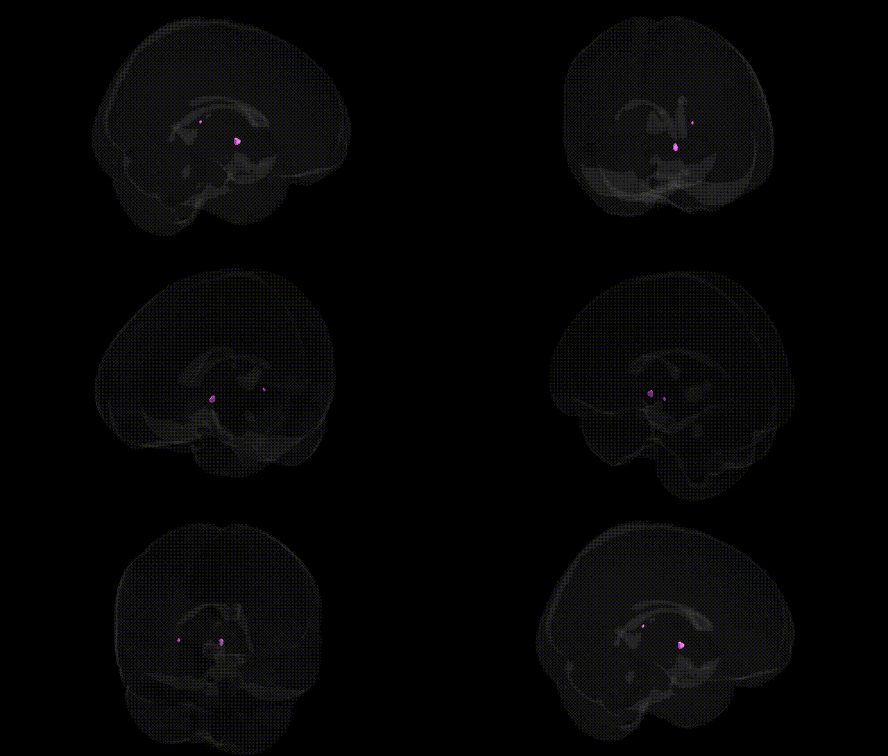
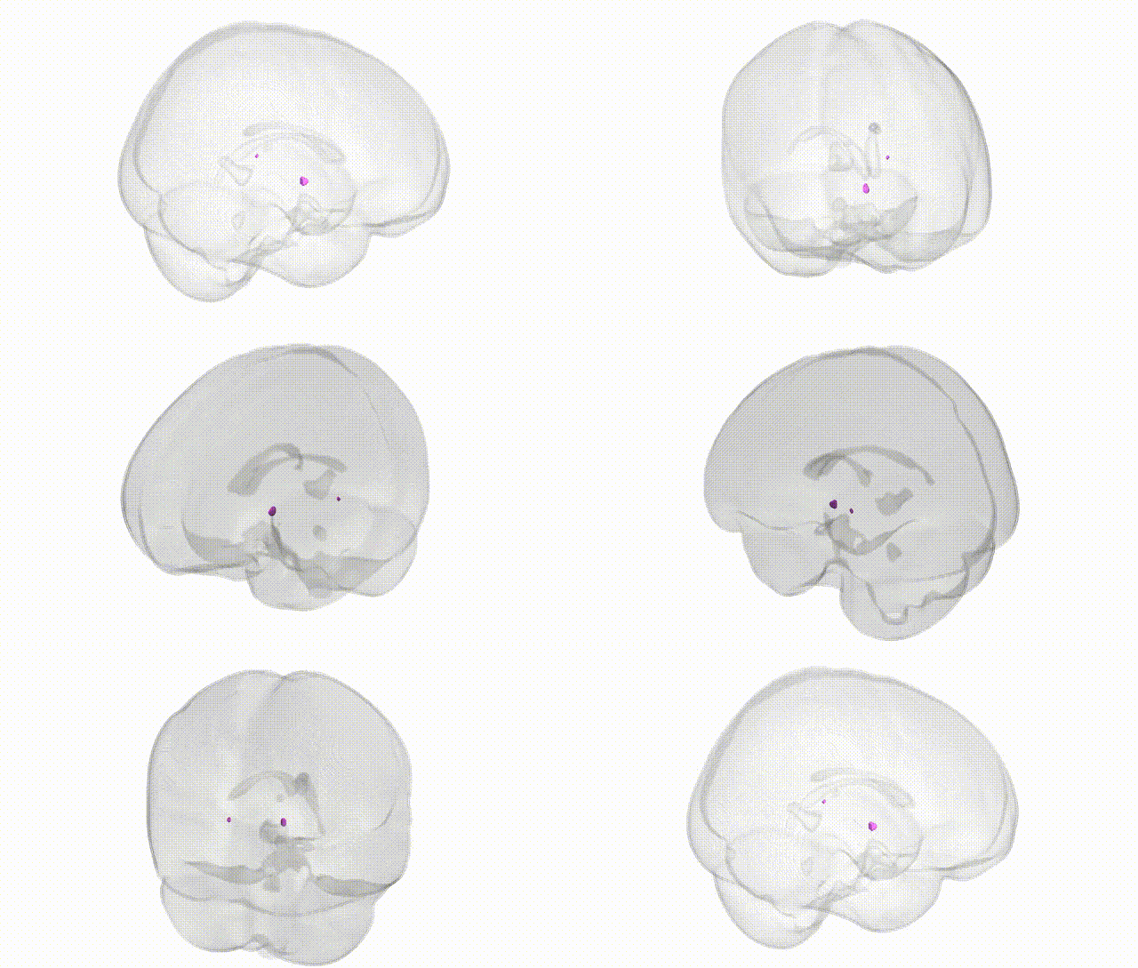
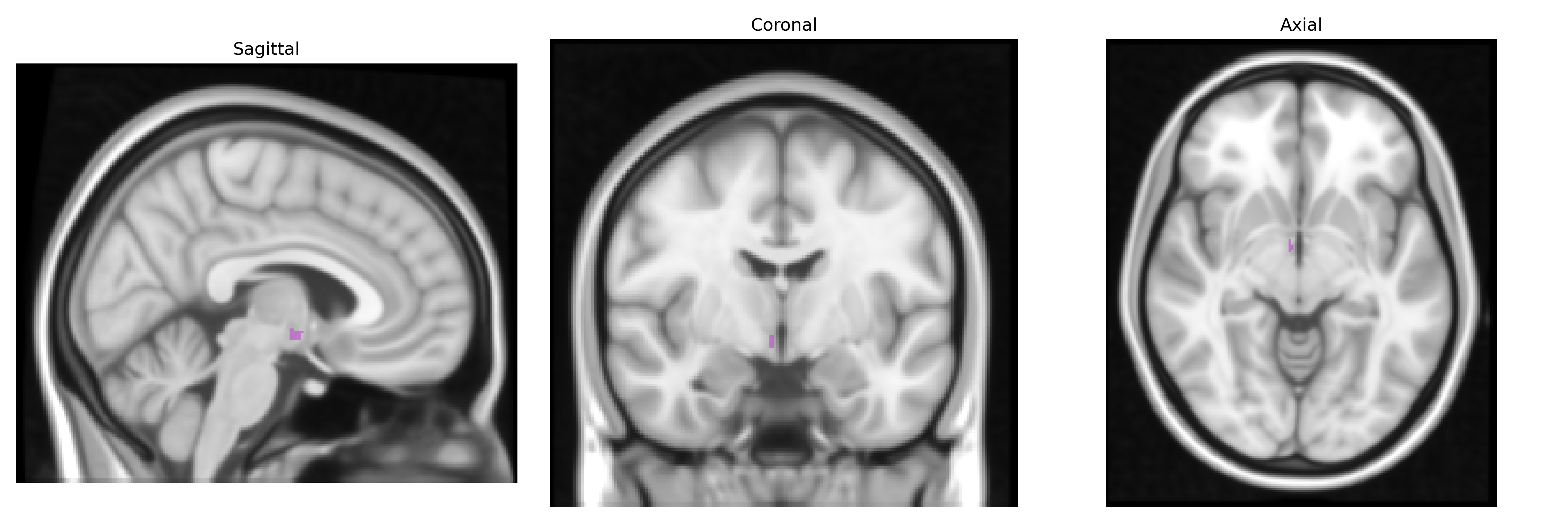
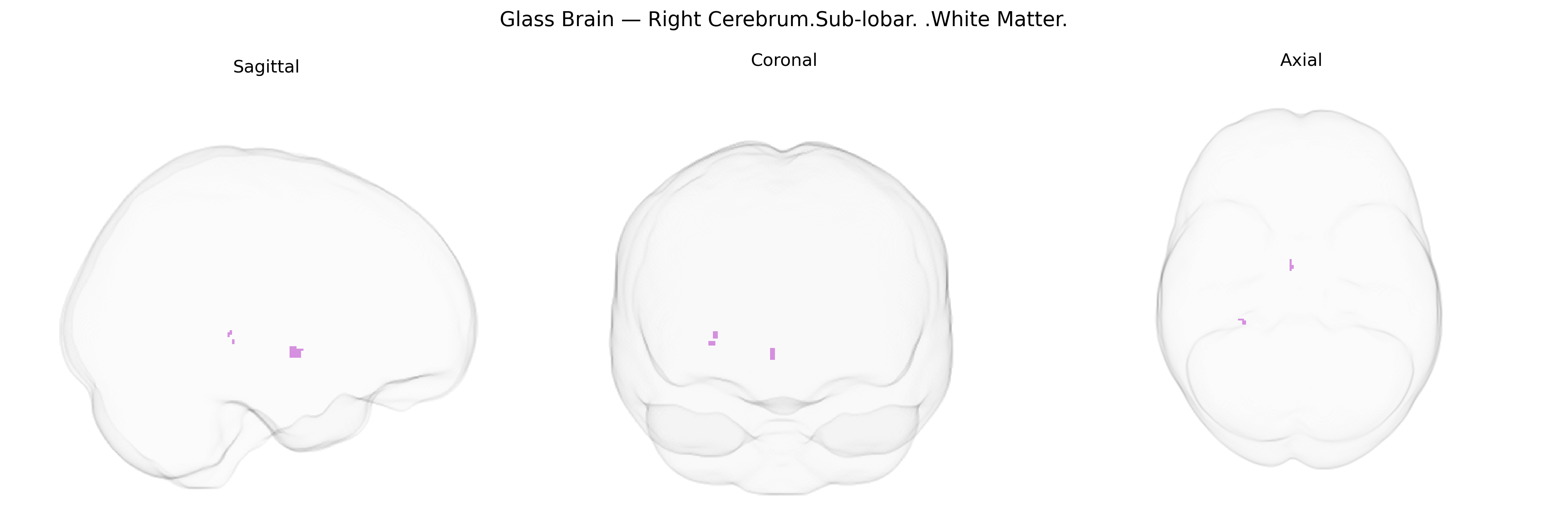
The bilateral Right Cerebrum Sub-lobar White Matter refers to the deep, non-cortical myelinated fiber tracts located beneath the sub-lobar cortical areas (such as insular and opercular regions) within the right cerebral hemisphere. This tissue consists primarily of association, commissural, and projection fibers that connect sub-lobar cortex with other cortical and subcortical structures, facilitating integration of sensory, motor, autonomic, and higher-order cognitive information across distributed neural networks. Functionally, these white matter pathways support inter-regional communication rather than local processing, enabling coordination between right-hemisphere sub-lobar regions (including insular and frontal opercular areas), the limbic system, basal ganglia, thalamus, and contralateral hemisphere via the corpus callosum. There is no direct link for this composite Talairach label; a closely related structure is the cerebral white matter: Cerebral white matter.
The bilateral right cerebrum sub‑lobar white matter, as defined in the Talairach 1 mm atlas, encompasses major intrahemispheric fiber tracts whose genetic architecture has been characterized primarily via diffusion MRI GWAS and large imaging‑genetics consortia (e.g., ENIGMA, UK Biobank). Common variants influencing global and regional white matter microstructure in this territory frequently map to genes involved in axon guidance, myelination, and oligodendrocyte biology, including loci near or within MAG, MBP, PLP1, LINGO1, and genes in the neuregulin–ErbB pathway, as well as broader neurodevelopmental regulators such as CNTNAP2 and NRXN1. Polygenic influences on white matter integrity in this region overlap with risk variants for schizophrenia, bipolar disorder, major depressive disorder, and autism spectrum conditions, consistent with findings that reduced fractional anisotropy and altered connectivity in right sub‑lobar white matter are enriched among carriers of psychiatric risk alleles. Additional GWAS links tie these white matter measures to variants associated with Alzheimer’s disease and other dementias (e.g., APOE, CLU, BIN1), ischemic stroke and small‑vessel disease (e.g., genes in the 17q25 locus such as TRIM47 and ACOX1), and neurodevelopmental traits including intelligence and educational attainment, indicating that genetic influences on this region’s white matter microstructure contribute to both normal cognitive variation and susceptibility to a range of neuropsychiatric and neurodegenerative disorders.
Overview generated by GPT-4o (2026).
Region ID: 462
Hemisphere: bilateral
Atlas: Talairach labels 1mm
Full Quality Version: Download MP4
Full Quality Version: Download MP4
Wali Sidiqyar*, Gaurav Rudravaram*, Elyssa M. McMaster, Trent M. Schwartz, Adam M. Saunders, Kurt G. Schilling, Bennett A. Landman "Introducing SPINS: A Shared Public Visualization Library of Neuroanatomical Structures." Medical Imaging with Deep Learning- short paper
This resource is licensed under CC0 1.0 Universal (Public Domain).