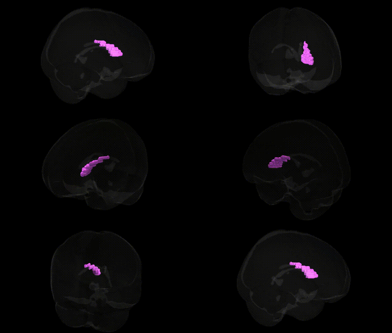
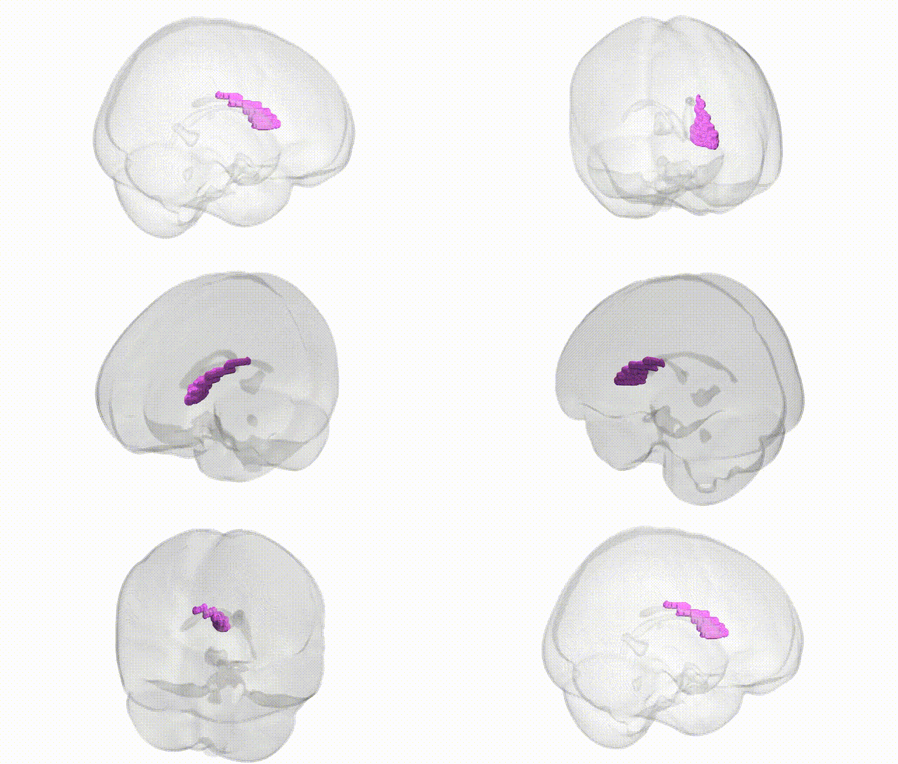
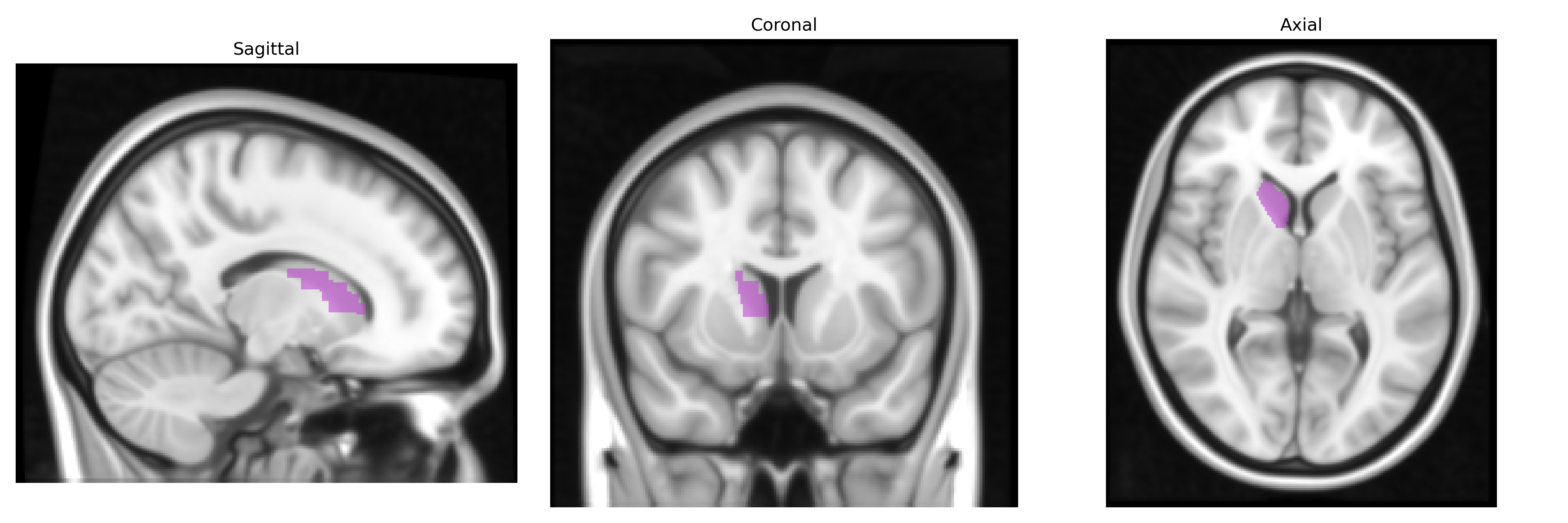
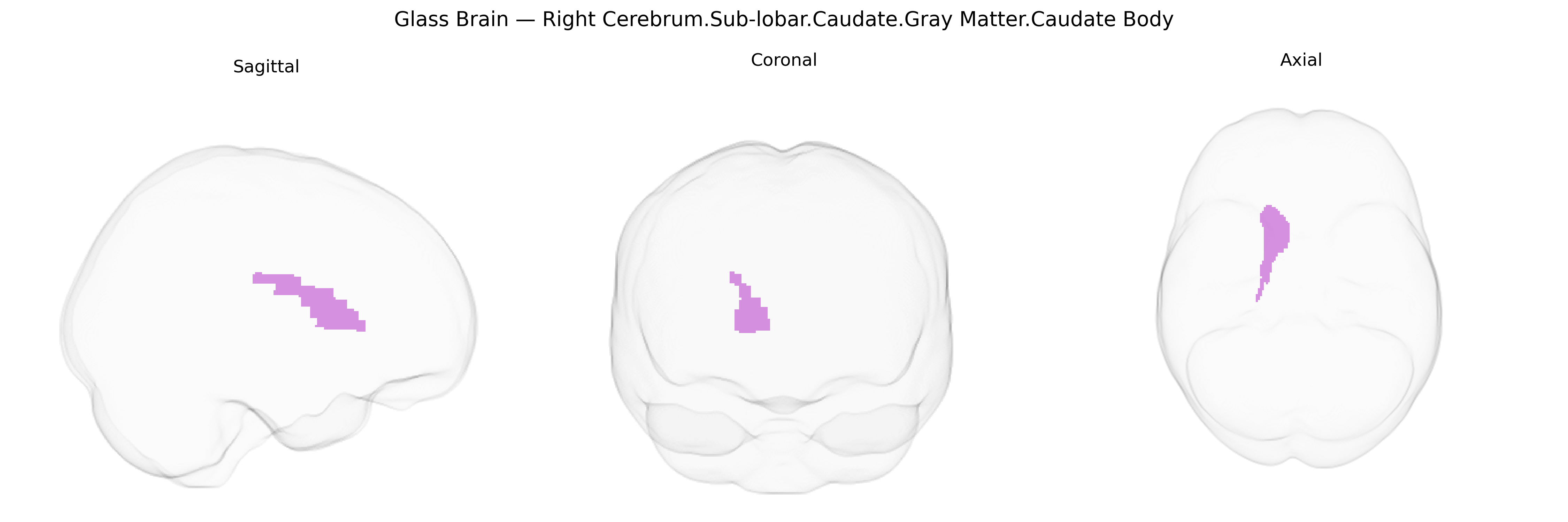
The bilateral Right Cerebrum.Sub-lobar.Caudate.Gray Matter.Caudate Body corresponds to the main elongated portion of the caudate nucleus, a C-shaped gray matter structure located deep within the subcortical (sub-lobar) region of the telencephalon. The caudate body, together with the caudate head and tail, forms part of the dorsal striatum and is a major input nucleus of the basal ganglia, receiving extensive glutamatergic projections from the cerebral cortex and dopaminergic input from the substantia nigra pars compacta. It plays a key role in motor control, procedural learning, action selection, habit formation, and aspects of cognition and motivation, integrating cortical information and influencing thalamocortical loops that modulate voluntary movement and goal-directed behavior. The caudate body lies lateral to the lateral ventricle and medial to the internal capsule, with its neurons organized into striosome and matrix compartments that participate in distinct basal ganglia circuits. Caudate nucleus
The bilateral caudate nucleus (Right Cerebrum.Sub-lobar.Caudate.Gray Matter.Caudate Body in the Talairach 1 mm atlas) has been implicated in multiple large-scale imaging genetics and GWAS studies, especially those quantifying subcortical volume and shape. Common variants in and near genes involved in neurodevelopment, synaptic function, and dopamine signaling—such as DRD2, COMT, BDNF, and several loci identified in ENIGMA and UK Biobank subcortical volume GWAS (e.g., variants near KNTC1, DCC, and other neurodevelopmental genes)—have shown associations with caudate size and morphology. Polygenic risk scores for schizophrenia, ADHD, obsessive–compulsive disorder, and major depression are associated with altered caudate volume and functional activity, and risk variants in genes like CACNA1C, GRIN2A, and others linked to glutamatergic and calcium-channel signaling have been related to striatal abnormalities that include the caudate. GWAS of cognitive traits, such as general intelligence, executive function, and educational attainment, frequently implicate genetic architectures that overlap with caudate volume–associated loci, consistent with the caudate’s role in fronto-striatal circuits supporting cognition and motivation. Additionally, genetic studies of Parkinson’s disease, Huntington’s disease (HTT expansions), and other movement disorders highlight the caudate as a key affected structure, with HTT CAG-repeat length and other neurodegenerative risk variants strongly associated with caudate atrophy, while substance use and addiction-related GWAS (e.g., for alcohol and nicotine dependence) also show convergent evidence of genetically influenced changes in caudate structure and function.
Overview generated by GPT-4o (2026).
Region ID: 693
Hemisphere: bilateral
Atlas: Talairach labels 1mm
Full Quality Version: Download MP4
Full Quality Version: Download MP4
Wali Sidiqyar*, Gaurav Rudravaram*, Elyssa M. McMaster, Trent M. Schwartz, Adam M. Saunders, Kurt G. Schilling, Bennett A. Landman "Introducing SPINS: A Shared Public Visualization Library of Neuroanatomical Structures." Medical Imaging with Deep Learning- short paper
This resource is licensed under CC0 1.0 Universal (Public Domain).