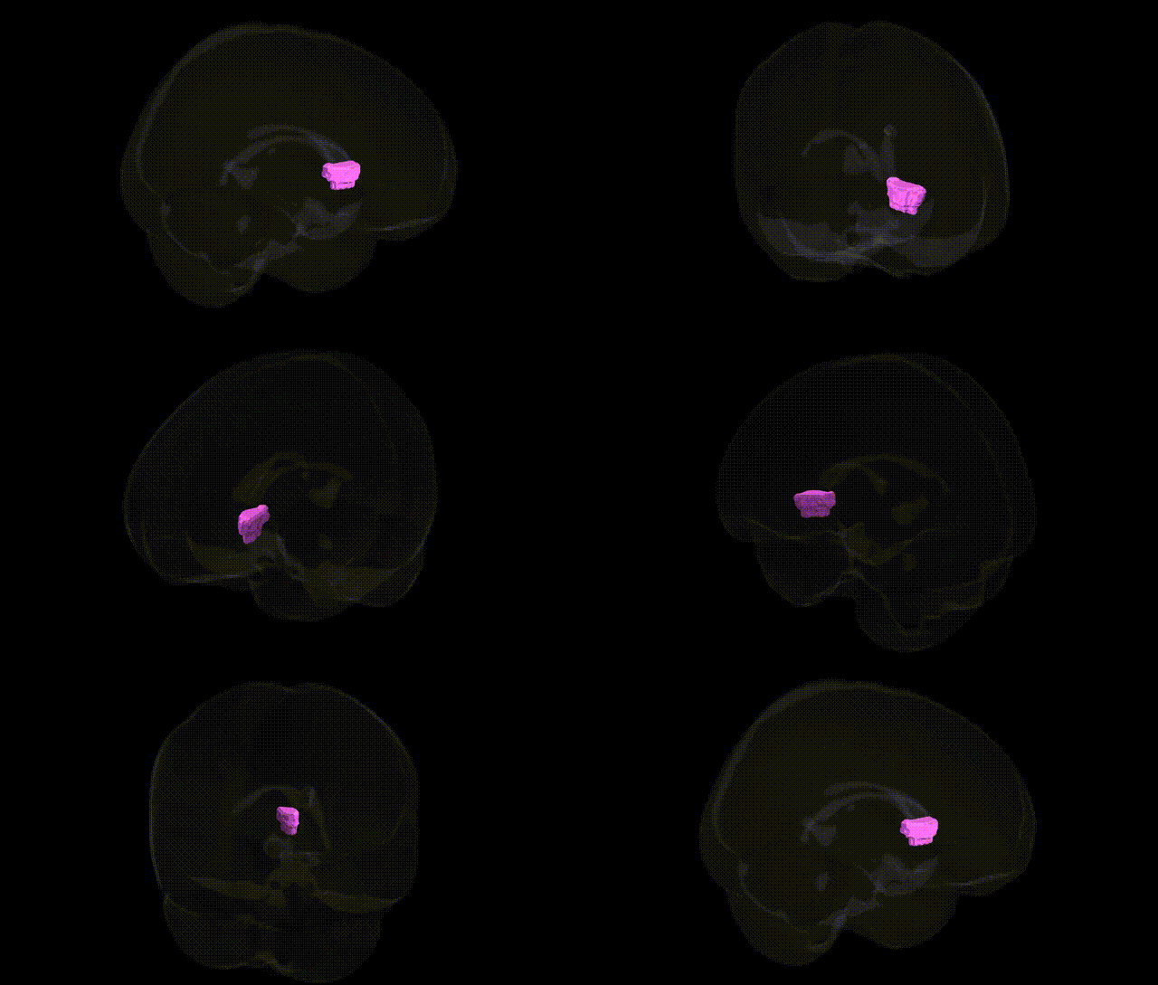
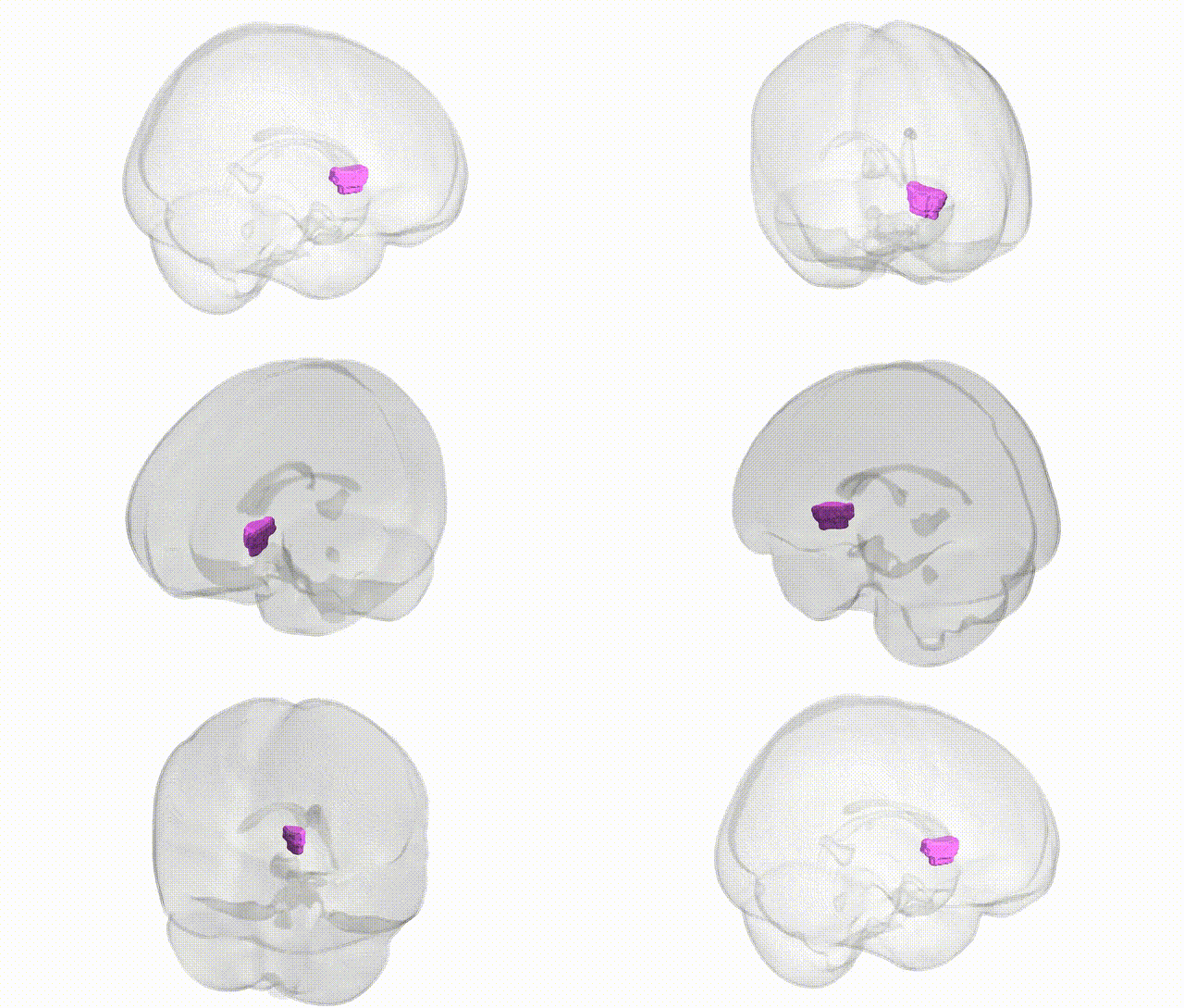
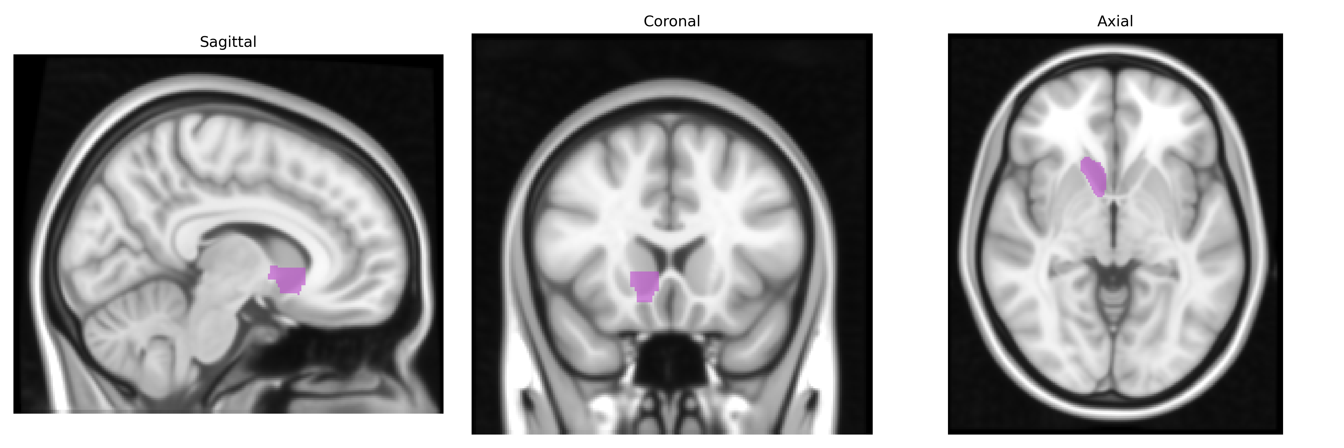
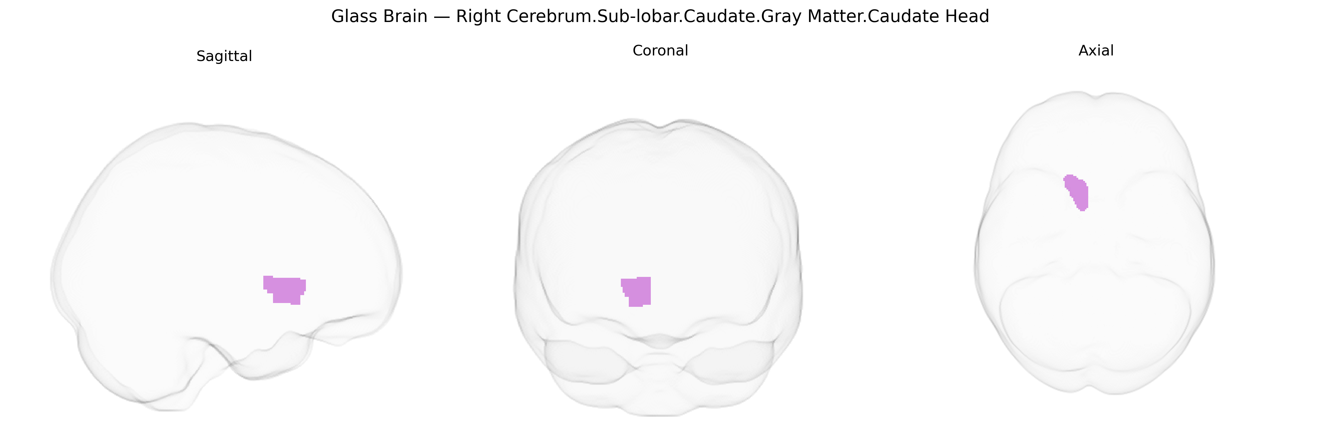
The bilateral Right Cerebrum.Sub-lobar.Caudate.Gray Matter.Caudate Head corresponds to the head of the caudate nucleus, a C-shaped gray matter structure situated in the subcortical (sub-lobar) region of the cerebral hemispheres, forming part of the dorsal striatum within the basal ganglia circuitry. It lies anteriorly and superiorly to the thalamus and lateral ventricle, with dense connections to the frontal cortex, especially prefrontal regions, and receives major dopaminergic input from the substantia nigra. Functionally, the caudate head is involved in motor control, procedural learning, action selection, reward-related processing, and aspects of cognition such as working memory and executive functions; it is also implicated in neuropsychiatric and movement disorders including Parkinson’s disease, Huntington’s disease, obsessive–compulsive disorder, and attention-deficit/hyperactivity disorder. There is no direct link specifically for “Caudate Head,” but it is a subregion of the Caudate Nucleus.
The bilateral caudate head gray matter in the sub-lobar right cerebrum, as defined in the Talairach 1 mm atlas, shows robust genetic influences on its volume and morphology, with SNP-based heritability typically in the 40–70% range across imaging–genetics studies such as ENIGMA and UK Biobank analyses. Large GWAS of subcortical volumes have identified multiple loci associated with caudate volume, including variants near genes involved in neurodevelopment and synaptic function (e.g., HMGA2, FAT3, DCC, and loci in or near WDR41 and LINC00461), and polygenic architecture overlapping with traits like cognitive ability and educational attainment. The caudate head is a key node in frontostriatal circuits, and genetic variation influencing this region has been implicated in neuropsychiatric disorders such as schizophrenia, bipolar disorder, major depressive disorder, ADHD, obsessive–compulsive disorder, Tourette syndrome, and autism spectrum disorder, where risk-associated variants often map to genes affecting dopaminergic signaling, synaptic plasticity, and neurodevelopment; in some cases, these risk alleles show parallel associations with altered caudate volume or functional activity in case-control imaging genetics. Neurodegenerative conditions such as Huntington’s disease (HTT CAG expansion) and, to a lesser degree, Parkinson’s disease and some forms of frontotemporal dementia are characterized by prominent caudate degeneration, and genetic risk for these disorders correlates with structural changes in the caudate head. Additionally, polygenic scores for metabolic and cardiometabolic traits (e.g., BMI, type 2 diabetes) and substance use have been linked to caudate structure and function, reflecting the region’s role in reward and habit learning, although many associations remain modest and highly polygenic.
Overview generated by GPT-4o (2026).
Region ID: 465
Hemisphere: bilateral
Atlas: Talairach labels 1mm
Full Quality Version: Download MP4
Full Quality Version: Download MP4
Wali Sidiqyar*, Gaurav Rudravaram*, Elyssa M. McMaster, Trent M. Schwartz, Adam M. Saunders, Kurt G. Schilling, Bennett A. Landman "Introducing SPINS: A Shared Public Visualization Library of Neuroanatomical Structures." Medical Imaging with Deep Learning- short paper
This resource is licensed under CC0 1.0 Universal (Public Domain).