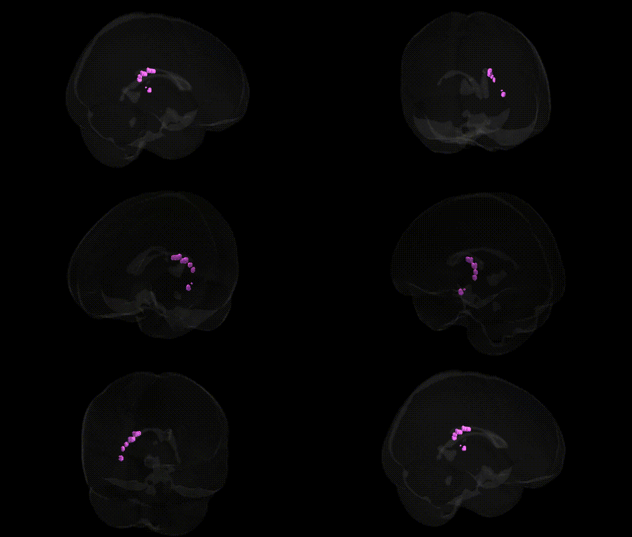
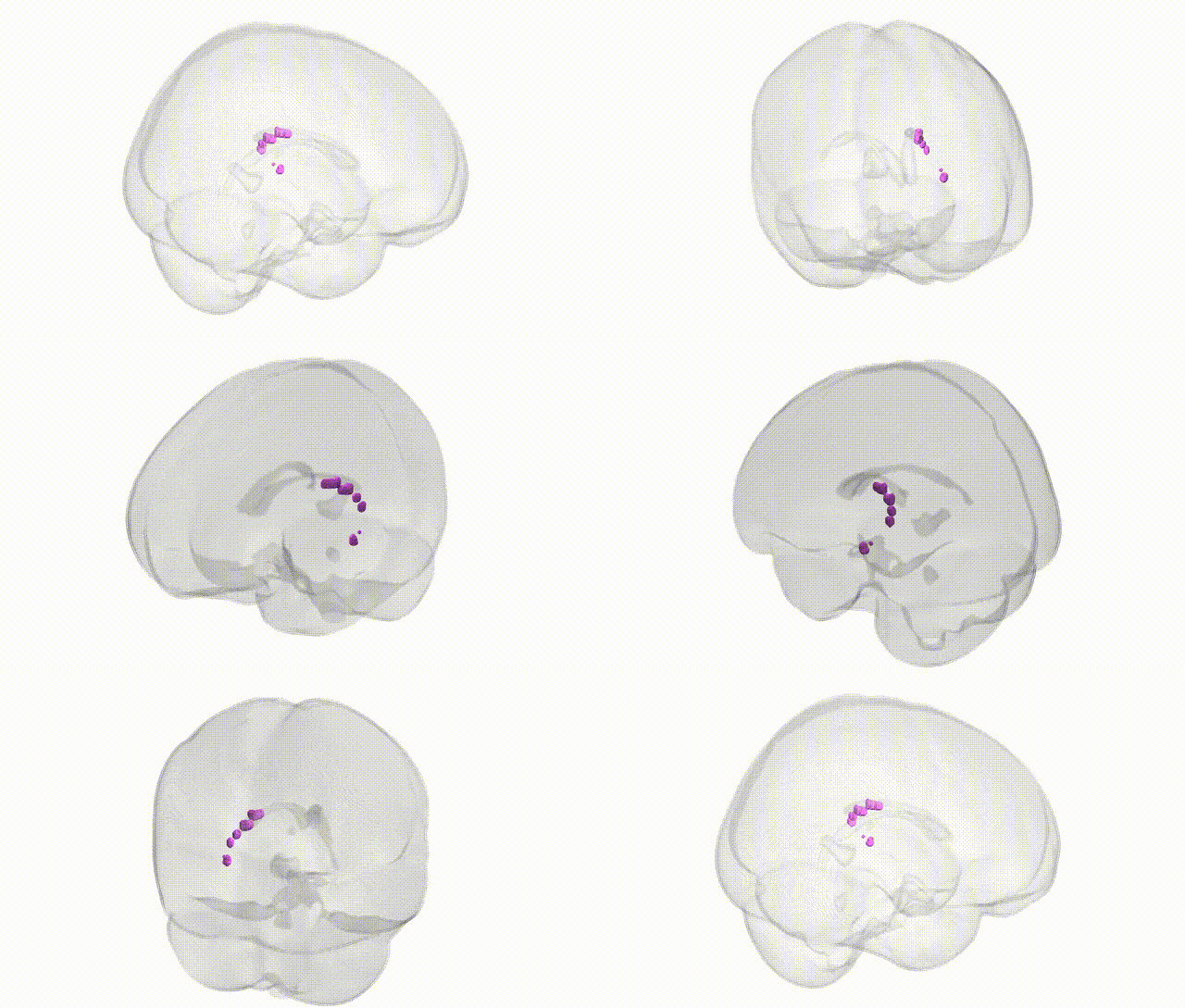
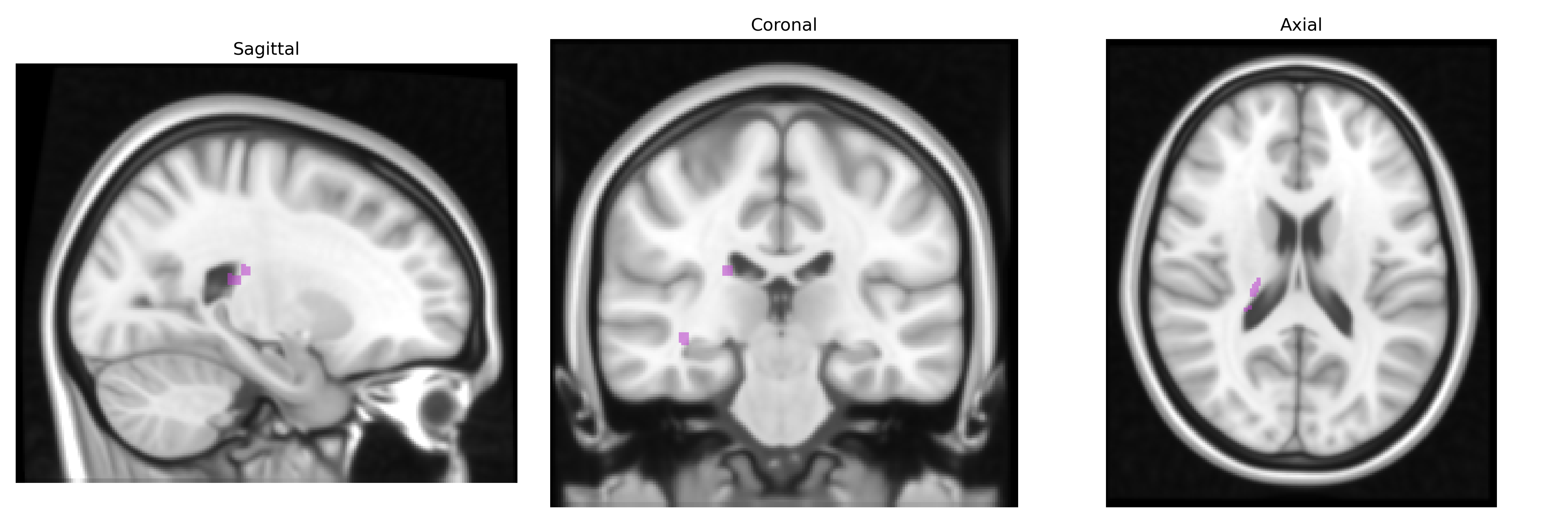
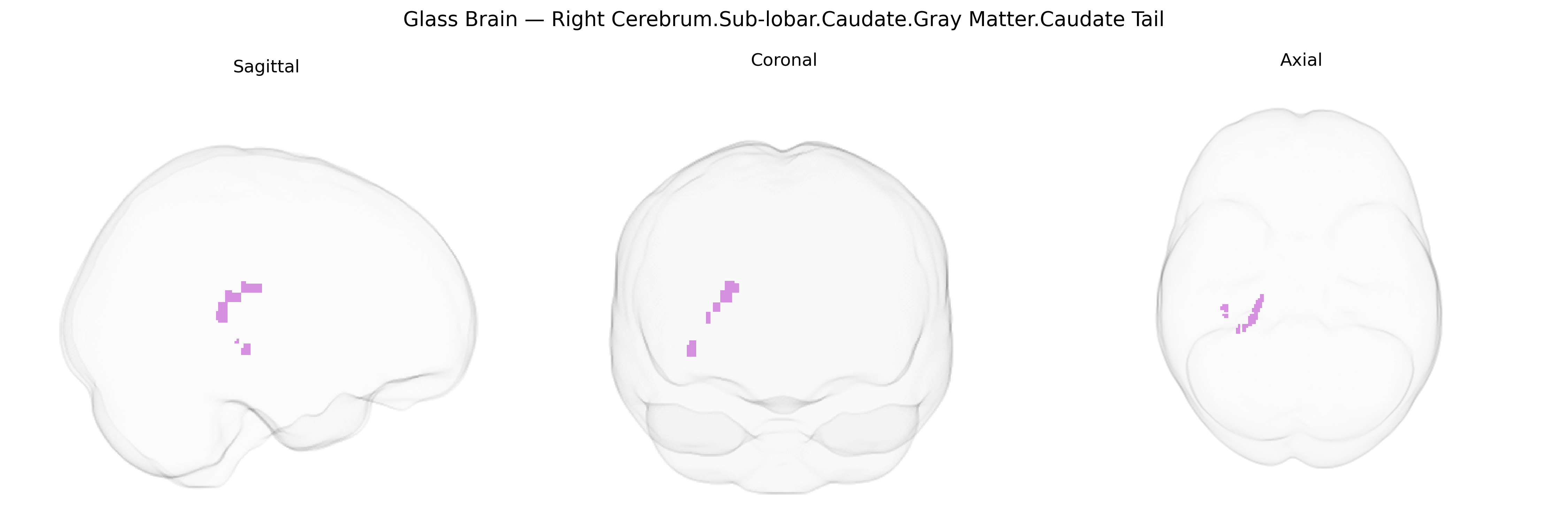
The bilateral Right Cerebrum.Sub-lobar.Caudate.Gray Matter.Caudate Tail region corresponds to the posterior portion of the caudate nucleus, a curved gray matter structure within the dorsal striatum of the basal ganglia. The caudate tail extends into the temporal lobe and lies adjacent to the lateral ventricle, participating in cortico-striato-thalamo-cortical loops. Functionally, this region is implicated in associative and limbic processing, including aspects of learning, memory, and emotional regulation, as well as integration of cognitive and motor information. It receives widespread cortical input and projects indirectly to motor and premotor areas via the globus pallidus and thalamus, contributing to the selection and gating of behavior. Pathological changes in caudate tail morphology or function have been associated with various neuropsychiatric and neurodegenerative conditions, including Huntington’s disease and obsessive-compulsive disorder. There is no direct link for the caudate tail; a closely related structure is the Caudate nucleus.
The caudate tail, a posterior segment of the caudate nucleus within the sublobar gray matter of the right cerebrum, has been implicated in multiple genetic associations largely inferred from imaging genetics and GWAS of striatal or caudate volume rather than this microanatomical subregion alone; common variants in genes related to dopamine signaling (e.g., DRD2, DAT1/SLC6A3), synaptic plasticity (e.g., BDNF), and neurodevelopment (e.g., NRG1, DISC1) have been associated with caudate structure and function, including alterations that extend into the tail, and polygenic risk scores for schizophrenia, bipolar disorder, and major depression have shown correlations with reduced or altered caudate volume in several large cohorts (e.g., ENIGMA). GWAS of subcortical volumes have identified loci near genes such as WDR41, DCC, and others involved in axon guidance and basal ganglia development that influence overall caudate morphology, likely affecting the tail region given its continuity with the caudate body, while copy number variants and rare coding mutations in neurodevelopmental genes (e.g., those implicated in autism spectrum disorder and intellectual disability) have been linked to atypical striatal development and connectivity that include the caudate tail’s visual and associative circuits. Genetic associations have also been reported between caudate measures and traits such as ADHD, obsessive-compulsive disorder, Tourette syndrome, and Parkinson’s disease, in which risk loci within dopaminergic, glutamatergic, and synaptic genes modulate basal ganglia-thalamo-cortical loops; additionally, variants influencing obesity, reward sensitivity, and addictive behaviors (e.g., FTO region, dopaminergic and opioid system genes) have been related to caudate-based reward circuitry that encompasses the tail. Collectively, current evidence suggests that genetic influences on the caudate tail are largely shared with those affecting the broader caudate and striatal network, with specific GWAS signals and risk alleles shaping its volume, connectivity, and role in neuropsychiatric and neurodevelopmental disorders, although fine-grained, tail-specific genetic mapping remains limited and is often inferred from higher-level caudate or striatal analyses rather than directly resolved in atlas-defined subregions such as the Talairach 1 mm caudate tail label.
Overview generated by GPT-4o (2026).
Region ID: 436
Hemisphere: bilateral
Atlas: Talairach labels 1mm
Full Quality Version: Download MP4
Full Quality Version: Download MP4
Wali Sidiqyar*, Gaurav Rudravaram*, Elyssa M. McMaster, Trent M. Schwartz, Adam M. Saunders, Kurt G. Schilling, Bennett A. Landman "Introducing SPINS: A Shared Public Visualization Library of Neuroanatomical Structures." Medical Imaging with Deep Learning- short paper
This resource is licensed under CC0 1.0 Universal (Public Domain).