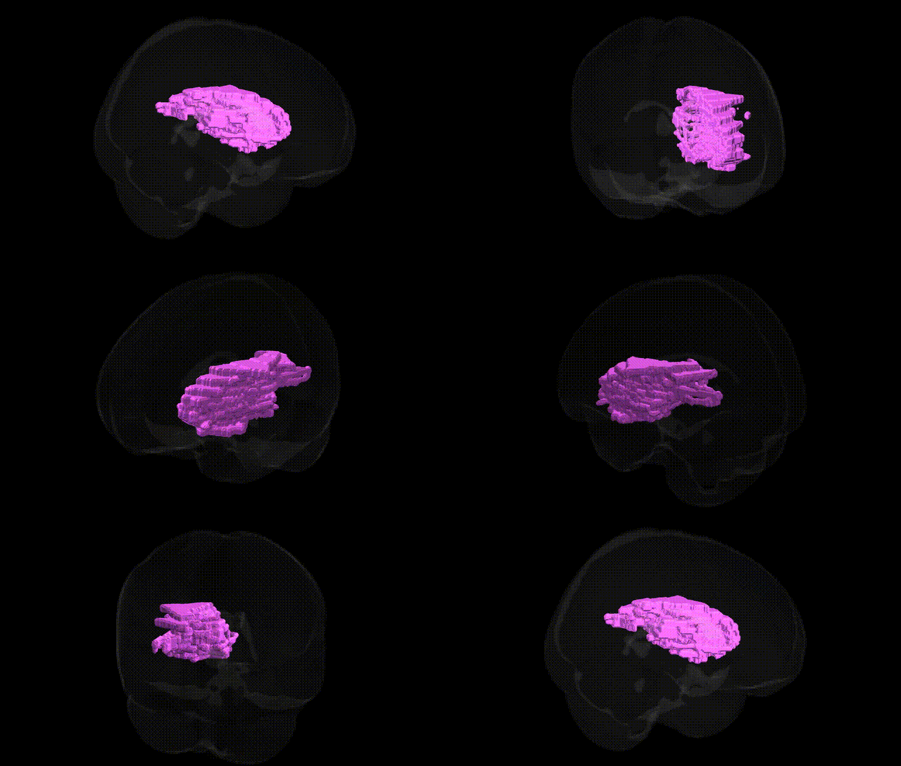
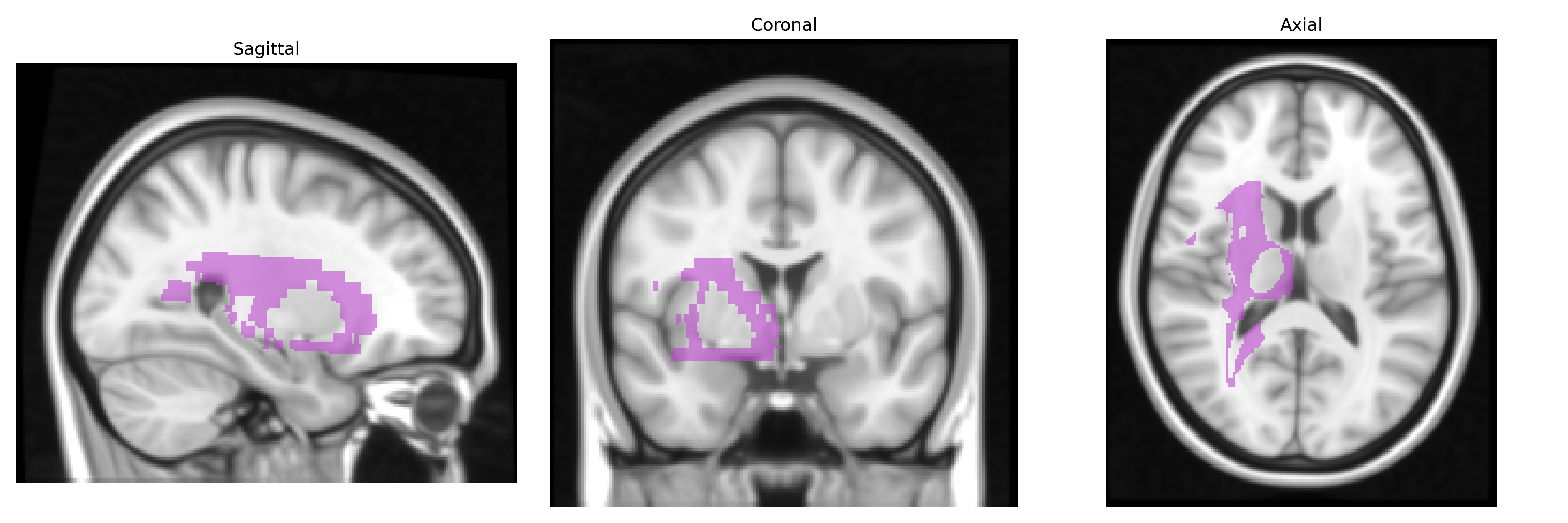
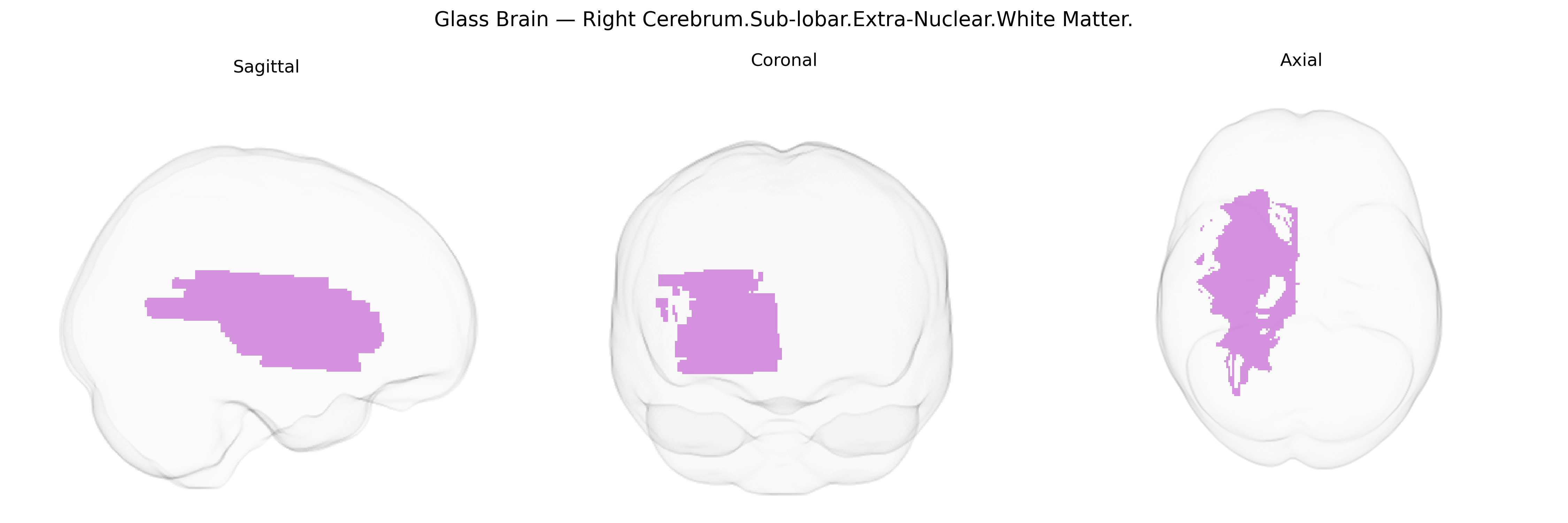
The bilateral Right Cerebrum.Sub-lobar.Extra-Nuclear.White Matter in the Talairach 1 mm Atlas refers to deep hemispheric white matter tracts located beneath the cortical mantle and outside (extra-) the major nuclear gray matter structures such as the basal ganglia and thalamus. This region comprises bundles of myelinated axons that interconnect cortical and subcortical areas, supporting long-range communication essential for motor control, sensory integration, cognition, and associative processing. It includes portions of major projection, association, and possibly commissural pathways coursing through the sublobar territory, helping coordinate information flow between frontal, parietal, temporal, and occipital regions as well as with deep nuclei and brainstem structures. There is no direct link for this exact label; a closely related structure is Cerebral white matter.
The bilateral Right Cerebrum.Sub-lobar.Extra-Nuclear.White Matter region, as defined in the Talairach 1 mm atlas, broadly corresponds to deep frontal and subcortical white matter tracts (e.g., frontostriatal and frontothalamic pathways), for which genetic associations are typically captured in large-scale imaging genetics studies examining white matter microstructure and connectivity rather than this specific atlas label. Genome-wide association studies (GWAS) of diffusion MRI measures (such as fractional anisotropy and mean diffusivity in frontal and subcortical white matter) have implicated loci near genes involved in myelination, axonal guidance, and oligodendrocyte biology, including but not limited to variants in or near NCAN, NTRK3, MAG, and genes regulating neuronal adhesion and cytoskeletal dynamics. These white matter tracts are frequently implicated as intermediate phenotypes in psychiatric and neurodevelopmental disorders such as schizophrenia, bipolar disorder, major depressive disorder, and ADHD, where polygenic risk scores correlate with altered integrity of frontal–subcortical white matter pathways. Additional associations arise in studies of cognitive traits (e.g., general intelligence, processing speed) and neurodegenerative or cerebrovascular conditions (Alzheimer’s disease, small vessel disease, leukoaraiosis), where risk variants in genes related to vascular function, inflammation, and myelin maintenance (for example, APOE and NOTCH3 in relevant contexts) have been linked to changes in deep frontal and extra-nuclear white matter. However, current GWAS and imaging-genetics findings do not typically resolve to the precise “Right Cerebrum.Sub-lobar.Extra-Nuclear.White Matter” Talairach label, so reported associations reflect broader deep frontal and subcortical white matter architecture rather than this specific atlas-defined parcel.
Overview generated by GPT-4o (2026).
Region ID: 348
Hemisphere: bilateral
Atlas: Talairach labels 1mm
Full Quality Version: Download MP4
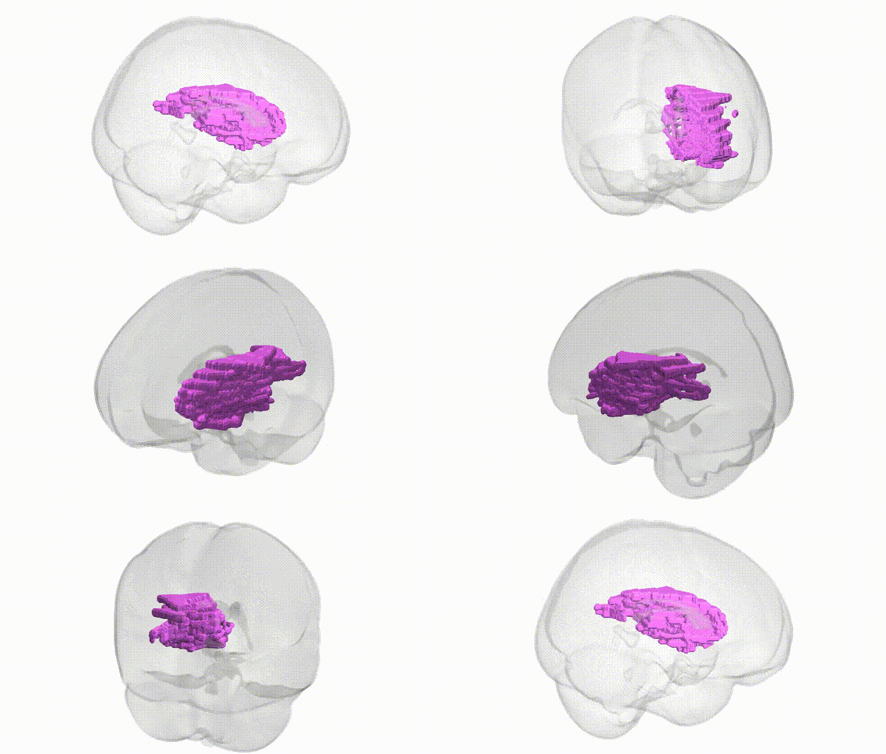
Full Quality Version: Download MP4
Wali Sidiqyar*, Gaurav Rudravaram*, Elyssa M. McMaster, Trent M. Schwartz, Adam M. Saunders, Kurt G. Schilling, Bennett A. Landman "Introducing SPINS: A Shared Public Visualization Library of Neuroanatomical Structures." Medical Imaging with Deep Learning- short paper
This resource is licensed under CC0 1.0 Universal (Public Domain).