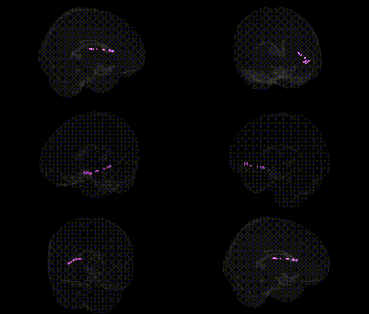
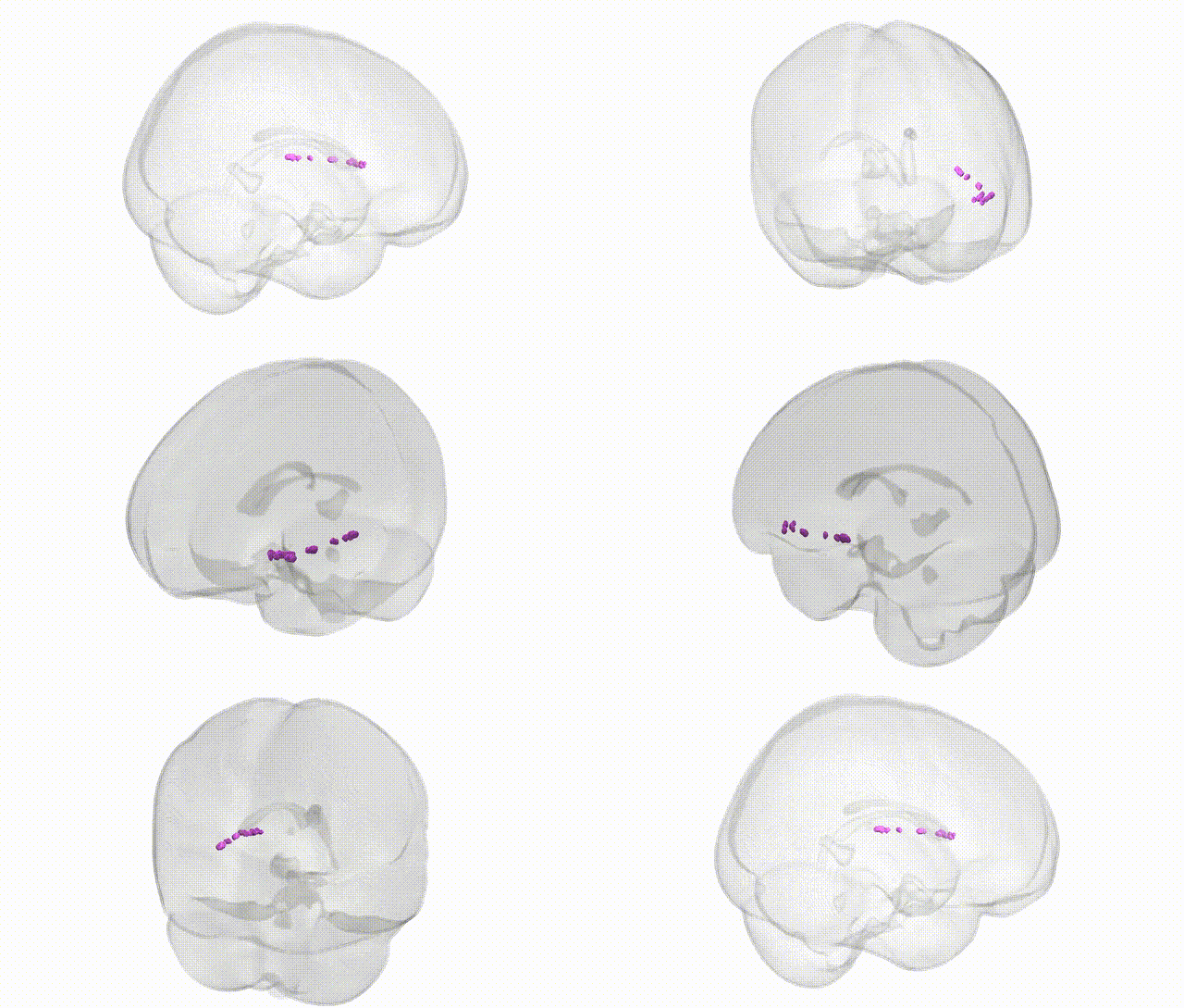
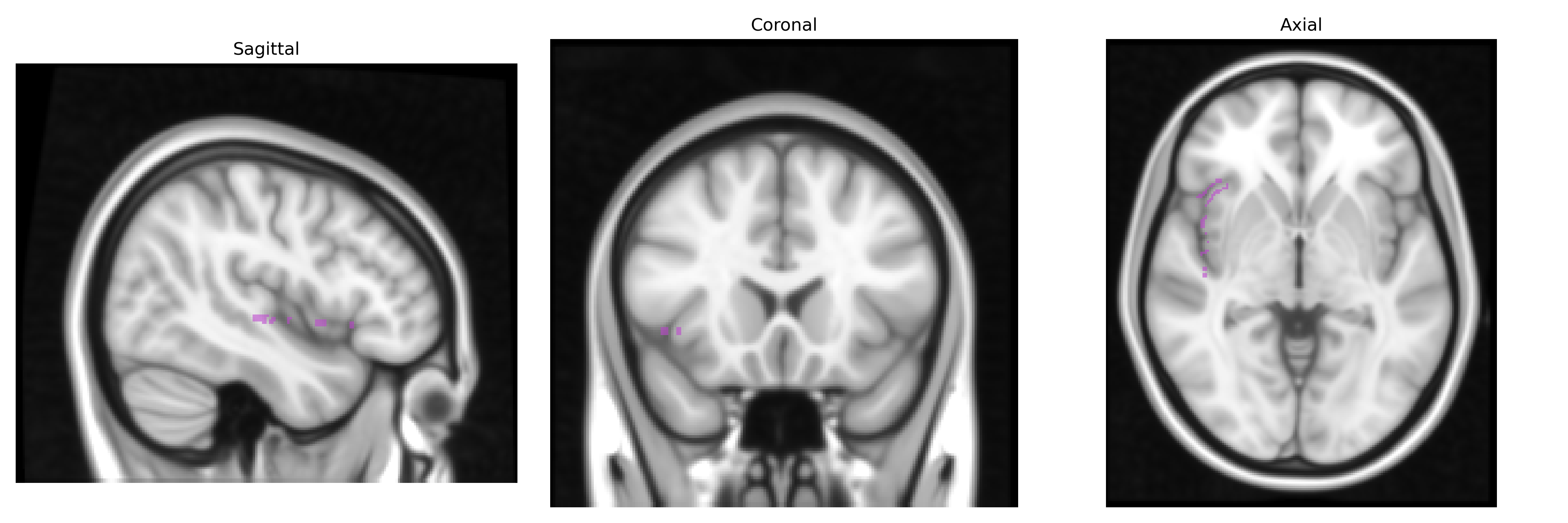
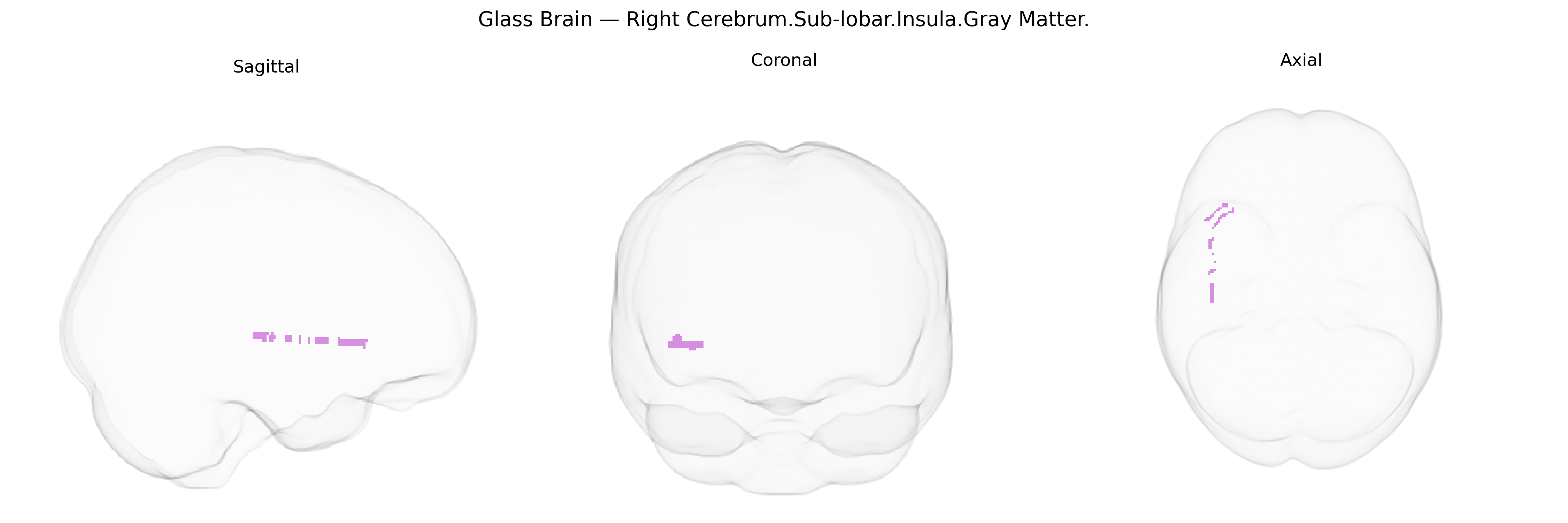
The bilateral Right Cerebrum.Sub-lobar.Insula.Gray Matter corresponds to the insular cortex, a deeply situated cortical region buried within the lateral sulcus beneath the frontal, parietal, and temporal opercula. This gray matter structure is involved in multimodal integration, including interoceptive awareness (perception of internal bodily states), gustatory processing, visceral sensation, pain perception, emotional and affective processing, and aspects of autonomic regulation. Functionally, the insula contributes to salience detection, integration of sensory and emotional information, and higher-order processes such as risk evaluation, empathy, and components of language and speech. Although asymmetries may exist between hemispheres, the bilateral insular cortices together form a key hub linking limbic, sensory, and cognitive networks. Insular cortex
The bilateral right insular cortex (Talairach “Right Cerebrum.Sub-lobar.Insula.Gray Matter”) has been implicated in multiple genetic and GWAS findings through its roles in emotion, interoception, and salience processing. Large neuroimaging-genetics consortia such as ENIGMA and UK Biobank have identified common variants (including loci near genes such as HMGA2, TESC, DCC, and others involved in neurodevelopment and synaptic function) associated with insular cortical thickness and surface area, suggesting heritable influences on its macrostructure. GWAS of psychiatric conditions—particularly schizophrenia, major depressive disorder, bipolar disorder, anxiety, and substance use disorders—have shown overlap between risk loci and regions whose morphology or functional connectivity prominently involve the insula, with polygenic risk scores for these disorders correlating with insular volume or activity changes in some studies. The insula also appears in genetic research on pain sensitivity, autonomic regulation, smoking and alcohol use, and obesity-related traits, where variants affecting dopaminergic, glutamatergic, and neuropeptide signaling show associations with altered insular responses or structure. In neurological disease, genes contributing to frontotemporal dementia, Alzheimer’s disease, and stroke risk have been linked to patterns of atrophy or functional disruption that prominently include the insula, reinforcing its role as a genetically influenced hub in networks underpinning salience, affective processing, and homeostatic regulation.
Overview generated by GPT-4o (2026).
Region ID: 585
Hemisphere: bilateral
Atlas: Talairach labels 1mm
Full Quality Version: Download MP4
Full Quality Version: Download MP4
Wali Sidiqyar*, Gaurav Rudravaram*, Elyssa M. McMaster, Trent M. Schwartz, Adam M. Saunders, Kurt G. Schilling, Bennett A. Landman "Introducing SPINS: A Shared Public Visualization Library of Neuroanatomical Structures." Medical Imaging with Deep Learning- short paper
This resource is licensed under CC0 1.0 Universal (Public Domain).