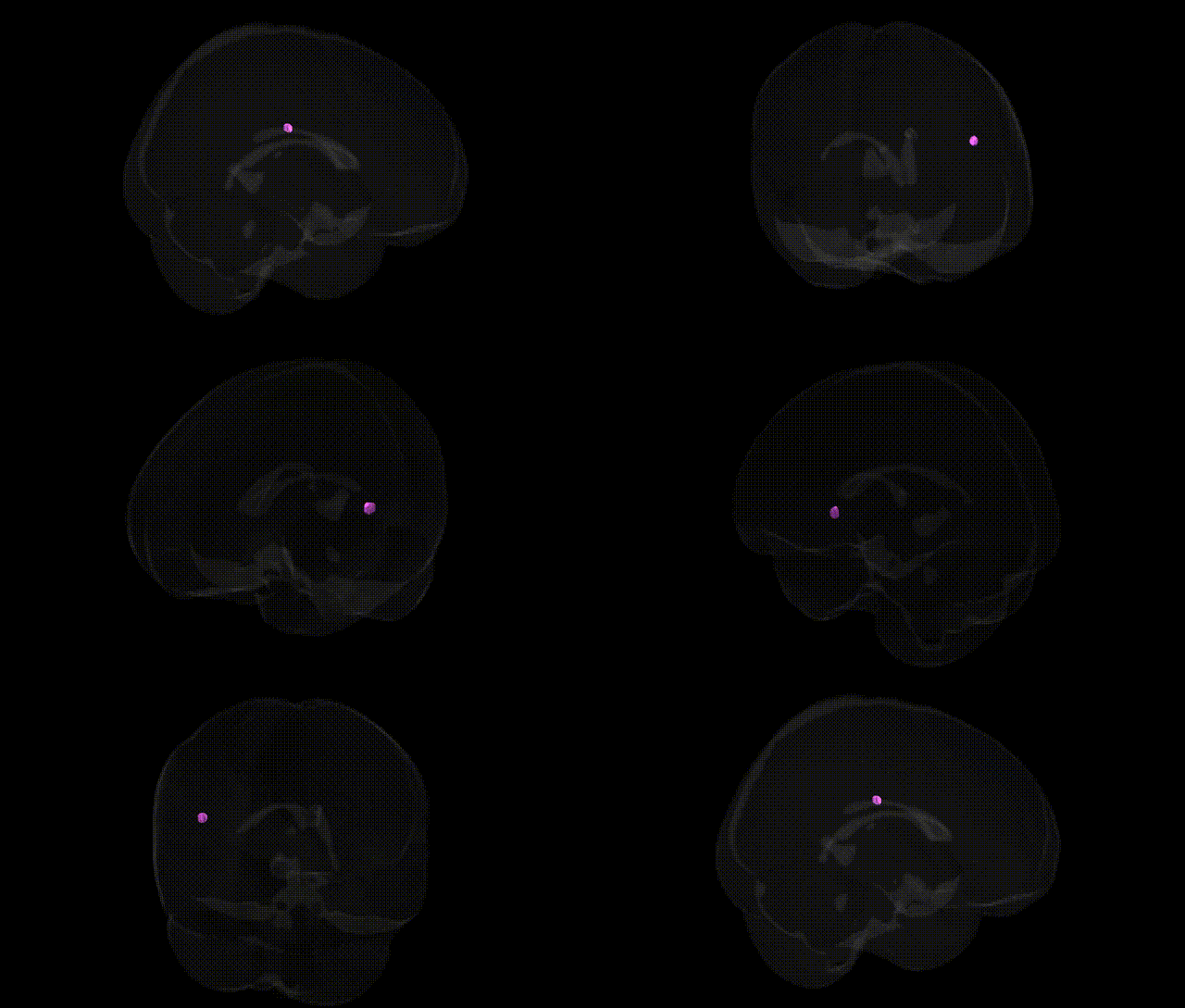
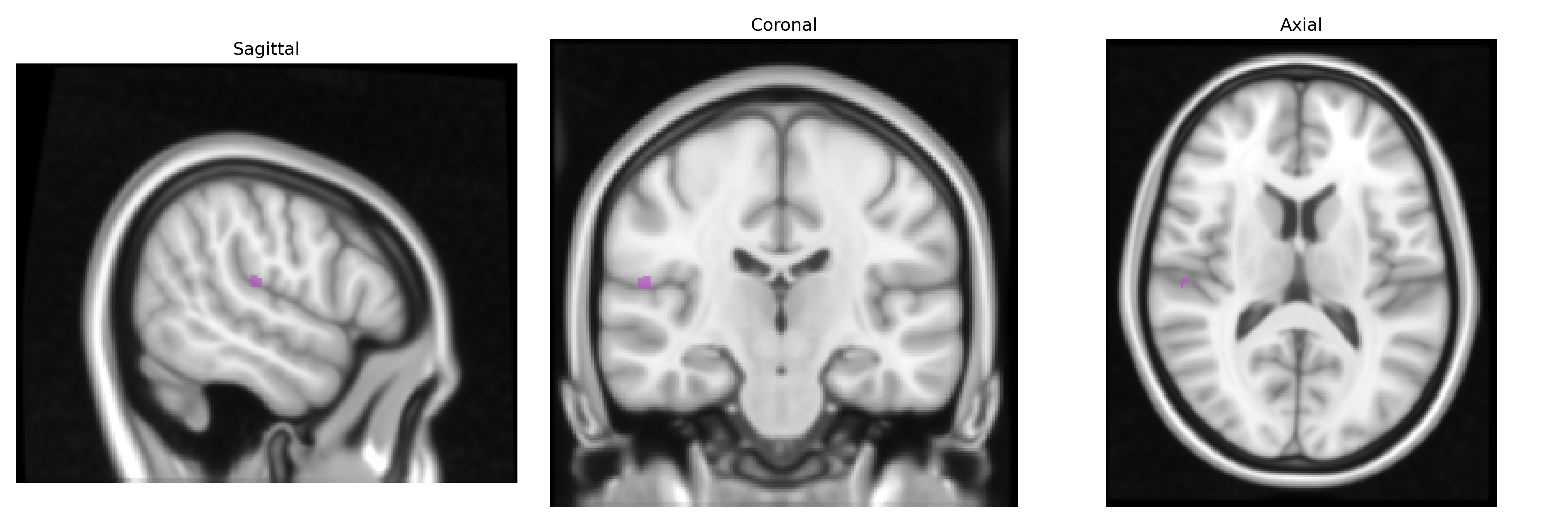
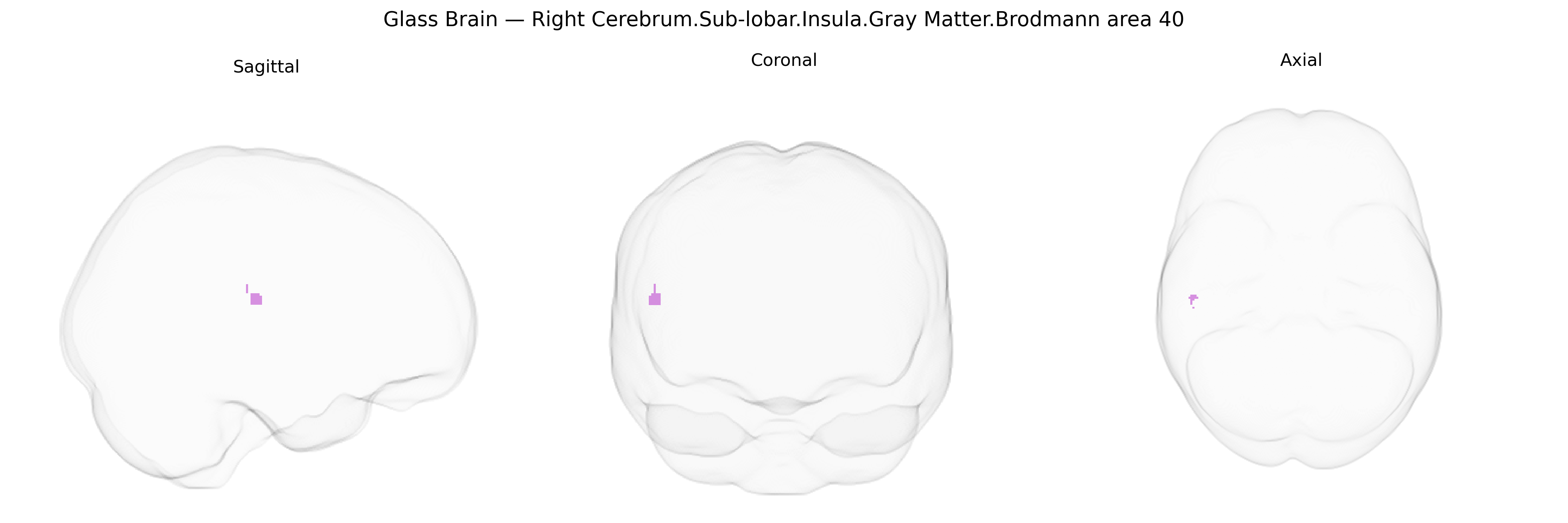
The bilateral Right Cerebrum.Sub-lobar.Insula.Gray Matter.Brodmann area 40 region, as labeled in the Talairach 1 mm Atlas, refers to parietal opercular cortex (classically supramarginal gyrus, Brodmann area 40) extending toward or overlapping the insular/opercular zone, situated deep to the lateral sulcus in the sub-lobar region of the right hemisphere and mirrored on the left. Functionally, this area is implicated in multimodal sensory integration, phonological and language-related processing, praxis, and aspects of spatial attention, as well as in higher-order somatosensory functions that interface with insular networks involved in interoception and salience detection. Although there is no dedicated Wikipedia article specifically for “Right Cerebrum.Sub-lobar.Insula.Gray Matter.Brodmann area 40,” a closely related structure and functional substrate is Brodmann area 40.
The right insular cortex, including sub-lobar gray matter regions spatially overlapping with Brodmann area 40 in Talairach-based atlases, shows multiple genetic associations in imaging-genetics and GWAS/meta-GWAS studies of cortical thickness, surface area, and volume, with heritability estimates often in the moderate range (h² ~0.3–0.6). Large consortia such as ENIGMA and UK Biobank–based analyses have identified common variants in genes related to neurodevelopment (e.g., microtubule and cytoskeletal genes, axon guidance, and synaptic signaling) that influence insular morphology, though findings are generally polygenic rather than region-specific and often overlap with nearby parietal/temporal association cortices. Genetic correlations link right insular structure with psychiatric and neurologic traits, including major depressive disorder, schizophrenia, bipolar disorder, autism spectrum disorder, and anxiety-related traits, as well as substance use phenotypes and pain sensitivity, consistent with the insula’s role in interoception, salience processing, and affect regulation. Some disease-focused GWAS and candidate-gene imaging studies have reported that risk alleles in genes such as CACNA1C, ZNF804A, and DISC1, among others, associate with altered insular volume or functional activation in patients and at-risk individuals, though effect sizes are small and findings not always replicated. In dementia and neurodegenerative conditions (e.g., Alzheimer’s disease, frontotemporal dementia), APOE and other risk variants show downstream associations with insular atrophy patterns in imaging-genetics analyses, again reflecting broad network-level vulnerability rather than a uniquely insula-specific genetic signal. Overall, genetic influences on this region appear highly polygenic and pleiotropic, shared with wider fronto-parietal and limbic networks rather than confined to a discrete Talairach-defined area.
Overview generated by GPT-4o (2026).
Region ID: 781
Hemisphere: bilateral
Atlas: Talairach labels 1mm
Full Quality Version: Download MP4
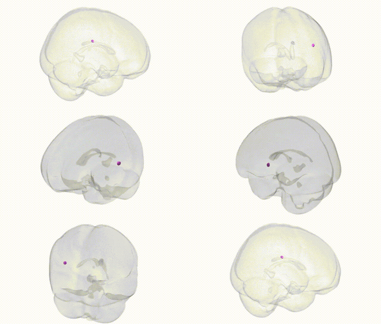
Full Quality Version: Download MP4
Wali Sidiqyar*, Gaurav Rudravaram*, Elyssa M. McMaster, Trent M. Schwartz, Adam M. Saunders, Kurt G. Schilling, Bennett A. Landman "Introducing SPINS: A Shared Public Visualization Library of Neuroanatomical Structures." Medical Imaging with Deep Learning- short paper
This resource is licensed under CC0 1.0 Universal (Public Domain).