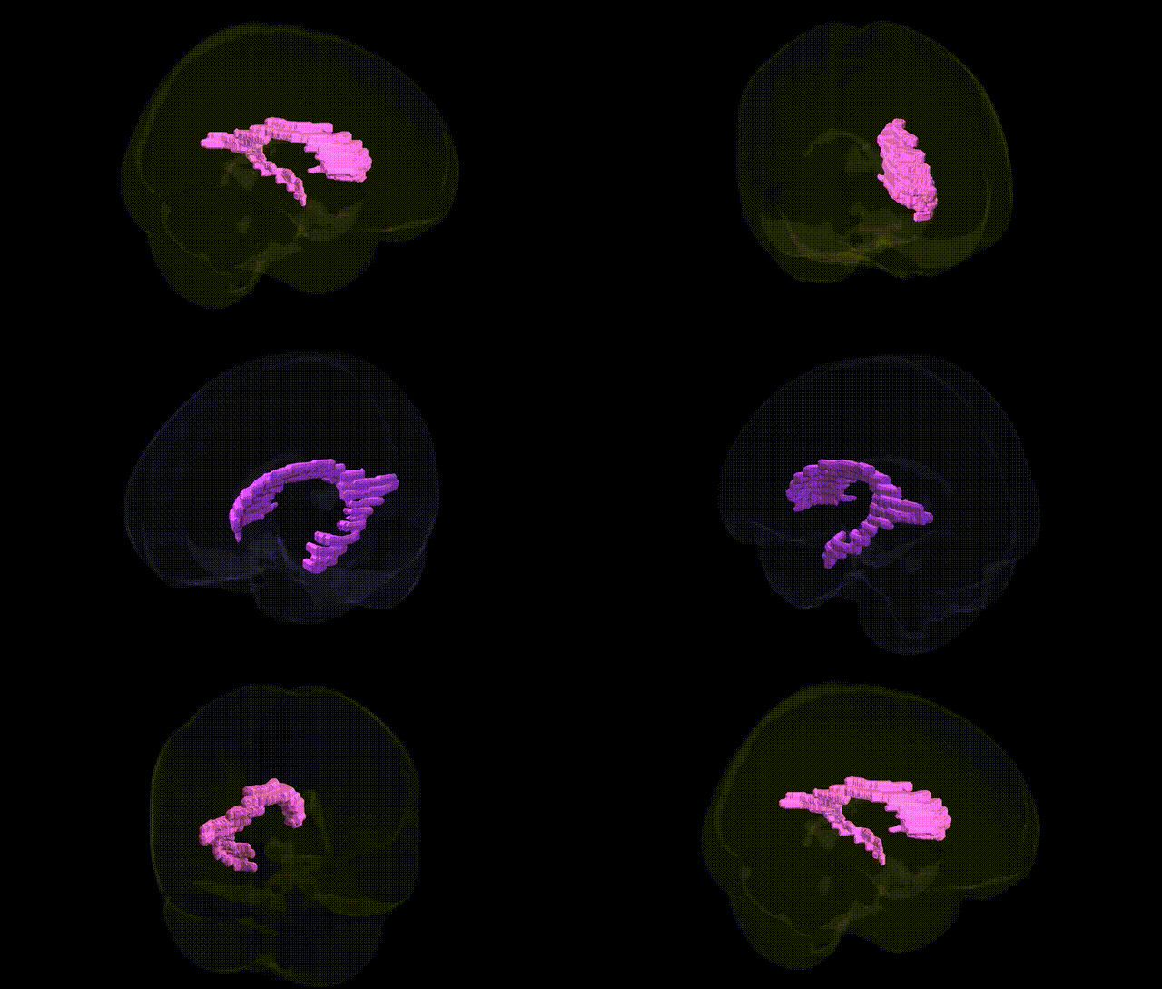
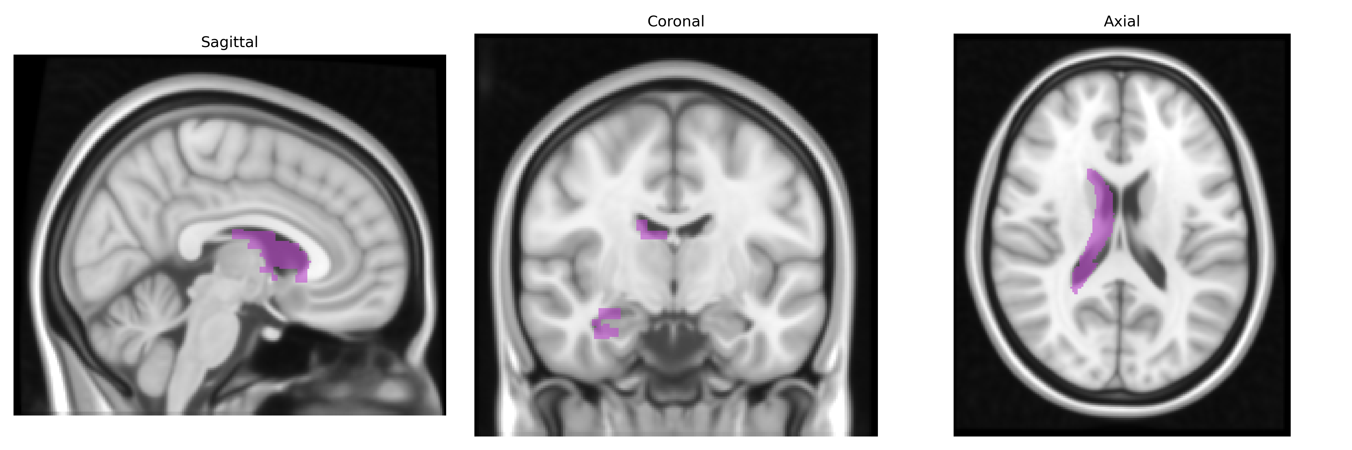
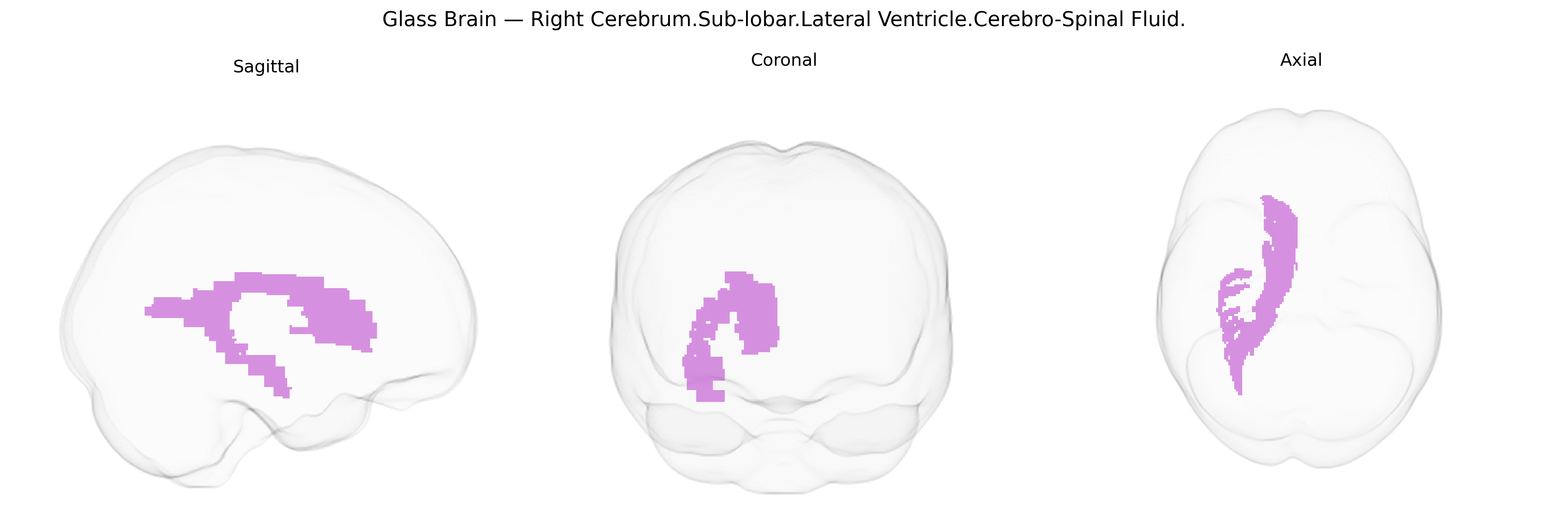
The bilateral Right Cerebrum.Sub-lobar.Lateral Ventricle.Cerebro-Spinal Fluid region corresponds to the cerebrospinal fluid (CSF)–filled cavity of the right lateral ventricle, a paired subcortical structure situated deep within the cerebral hemispheres. The lateral ventricle follows the contours of the surrounding lobes (frontal, parietal, temporal, and occipital) and is divided into anterior, posterior, inferior horns, and a central body. It is lined by ependymal cells and is part of the ventricular system that circulates CSF, which cushions the brain, maintains intracranial pressure, and contributes to metabolic waste removal. The “sub-lobar” designation reflects its position beneath the cortical mantle, adjacent to structures such as the caudate nucleus, corpus callosum, and thalamus. CSF in this region is produced primarily by the choroid plexus and communicates with other ventricles, playing a crucial role in normal brain homeostasis and neuroimaging contrast.
Genetic associations for the bilateral Right Cerebrum.Sub-lobar.Lateral Ventricle.Cerebro-Spinal Fluid region, as defined in the Talairach 1 mm atlas, emerge primarily from genome-wide association studies of ventricular and global brain volumetric measures rather than region-specific functional studies. Numerous large GWAS consortia (e.g., ENIGMA, UK Biobank–based studies) have identified common variants in genes such as APOE, MAPT, and loci near WNT3, GMNC, and LRRK2 that influence lateral ventricle volume, often interpreted as markers of neurodegeneration, brain atrophy, or neurodevelopmental variation. Increased lateral ventricle cerebrospinal fluid volume has been genetically linked to risk or progression of disorders including Alzheimer’s disease, schizophrenia, bipolar disorder, and other neuropsychiatric conditions, with shared polygenic architecture between ventricle size and these traits. Many loci overlap with those affecting global or regional cortical thickness, subcortical volumes, and white matter integrity, indicating that genetic influences on ventricular CSF largely reflect generalized brain structural vulnerability rather than lateral ventricle–specific biology.
Overview generated by GPT-4o (2026).
Region ID: 174
Hemisphere: bilateral
Atlas: Talairach labels 1mm
Full Quality Version: Download MP4
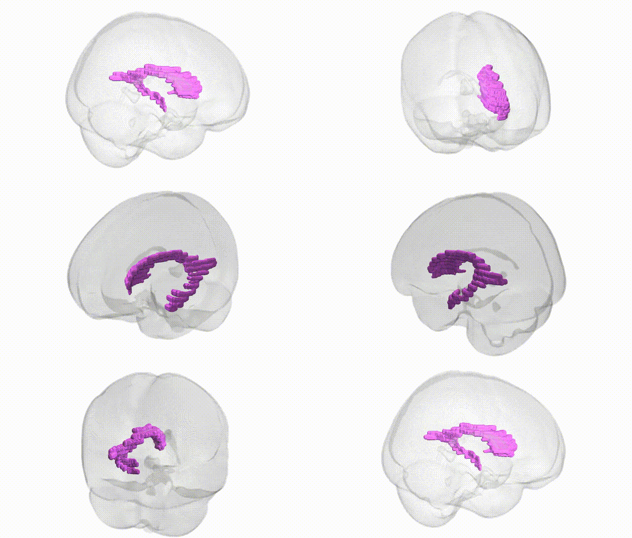
Full Quality Version: Download MP4
Wali Sidiqyar*, Gaurav Rudravaram*, Elyssa M. McMaster, Trent M. Schwartz, Adam M. Saunders, Kurt G. Schilling, Bennett A. Landman "Introducing SPINS: A Shared Public Visualization Library of Neuroanatomical Structures." Medical Imaging with Deep Learning- short paper
This resource is licensed under CC0 1.0 Universal (Public Domain).