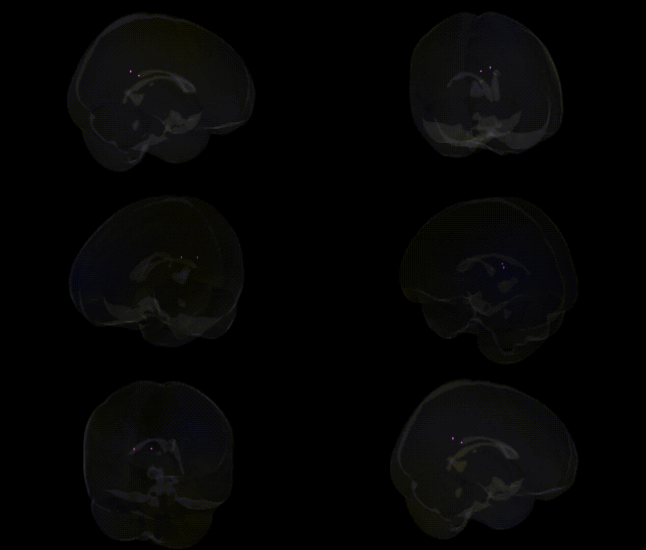
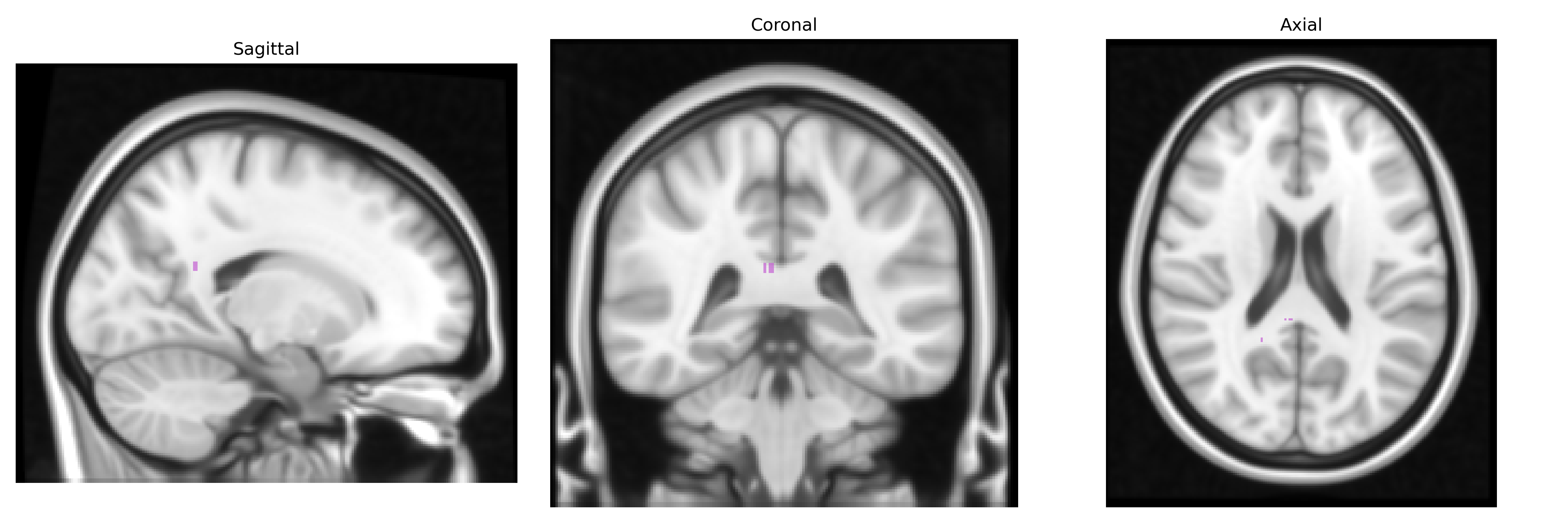
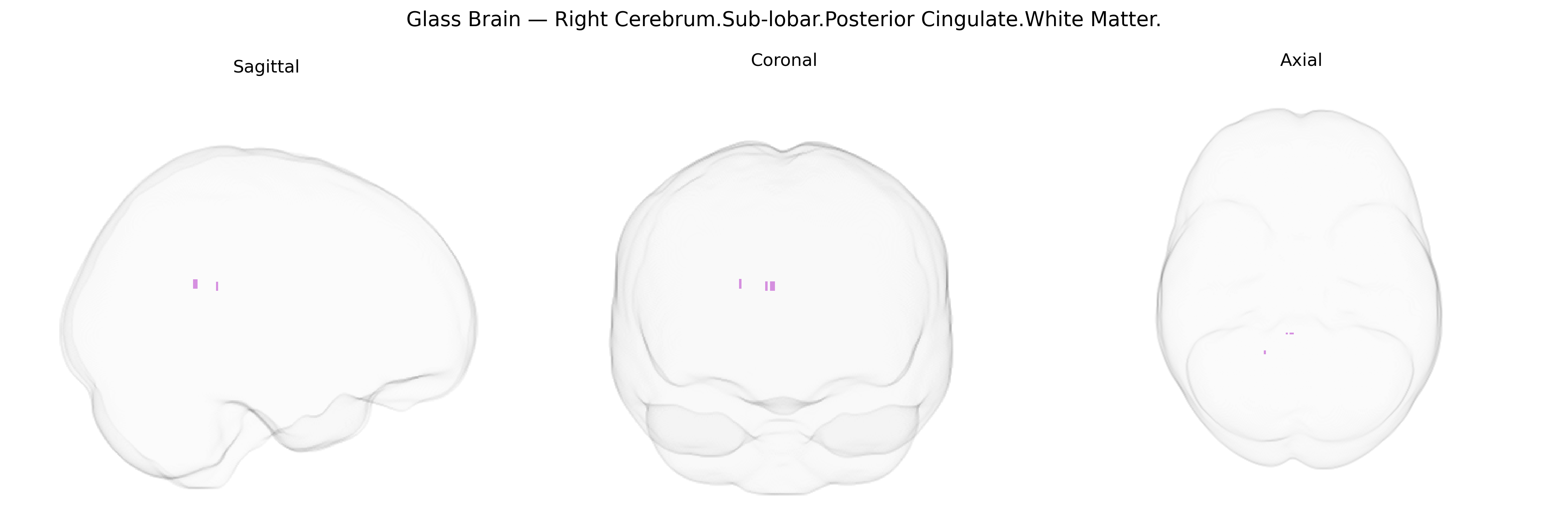
The bilateral Right Cerebrum.Sub-lobar.Posterior Cingulate.White Matter corresponds to the deep intrahemispheric fiber pathways underlying the posterior cingulate cortex, a key hub of the limbic and default mode networks. These white matter tracts connect the posterior cingulate gyrus with neighboring cingulate regions, the precuneus, medial parietal cortex, and other associative and limbic areas, supporting integration of self-referential thought, internally directed attention, episodic memory, and aspects of visuospatial and emotional processing. Damage or disruption of these fibers is associated with alterations in consciousness, memory impairments, and changes in default mode network connectivity observed in conditions such as Alzheimer’s disease and other neurodegenerative or demyelinating disorders. There is no direct link for this exact white matter label; a closely related cortical region is the Posterior cingulate cortex.
The bilateral right sublobar posterior cingulate white matter, as defined in the Talairach 1 mm atlas, lies within the broader posterior cingulate cortex (PCC) / cingulum bundle network, for which genetic associations are better characterized at the regional and connectivity level than at fine-grained atlas parcels. Large neuroimaging GWAS consortia (e.g., ENIGMA, UK Biobank) have identified multiple common variants influencing posterior cingulate cortical thickness, surface area, and white matter microstructure (often indexed via cingulum fractional anisotropy), implicating genes involved in neurodevelopment, myelination, and synaptic function such as those in the MAPT region on 17q21, GRIN2B, and several loci near oligodendrocyte- and axon-related genes. These structural and connectivity measures in posterior cingulate/cingulum pathways show genetic correlations with Alzheimer’s disease, general cognitive ability, and educational attainment, as well as with psychiatric conditions including major depressive disorder, schizophrenia, and bipolar disorder, consistent with the PCC’s role in default mode network function and memory. APOE ε4 and other Alzheimer’s risk loci show associations with posterior cingulate atrophy and white matter changes, while polygenic risk scores for dementia and psychiatric disorders have been linked to altered PCC structure and connectivity, suggesting that genetic risk for neurodegeneration and psychopathology may be partially mediated through this region’s white matter integrity.
Overview generated by GPT-4o (2026).
Region ID: 832
Hemisphere: bilateral
Atlas: Talairach labels 1mm
Full Quality Version: Download MP4
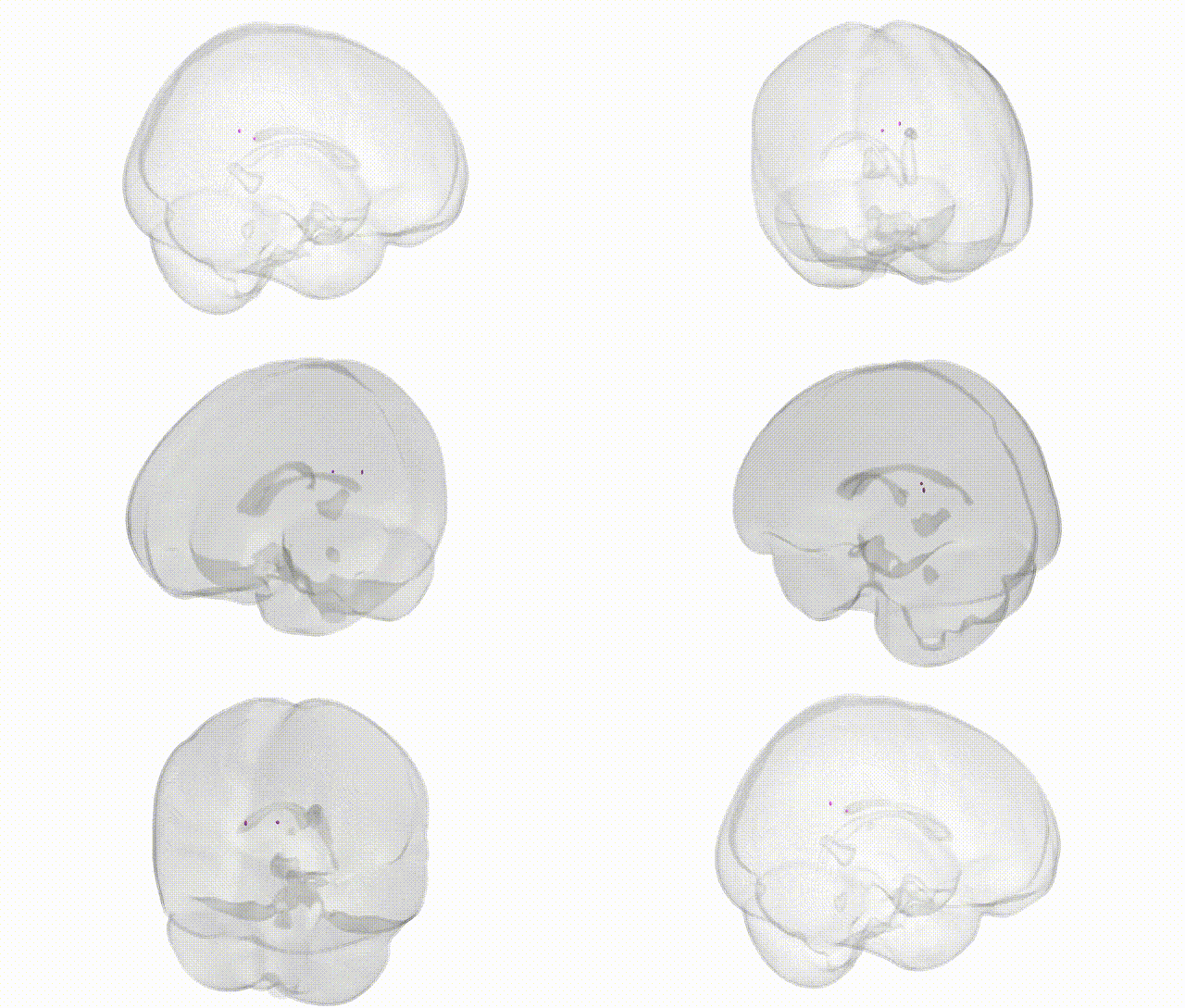
Full Quality Version: Download MP4
Wali Sidiqyar*, Gaurav Rudravaram*, Elyssa M. McMaster, Trent M. Schwartz, Adam M. Saunders, Kurt G. Schilling, Bennett A. Landman "Introducing SPINS: A Shared Public Visualization Library of Neuroanatomical Structures." Medical Imaging with Deep Learning- short paper
This resource is licensed under CC0 1.0 Universal (Public Domain).