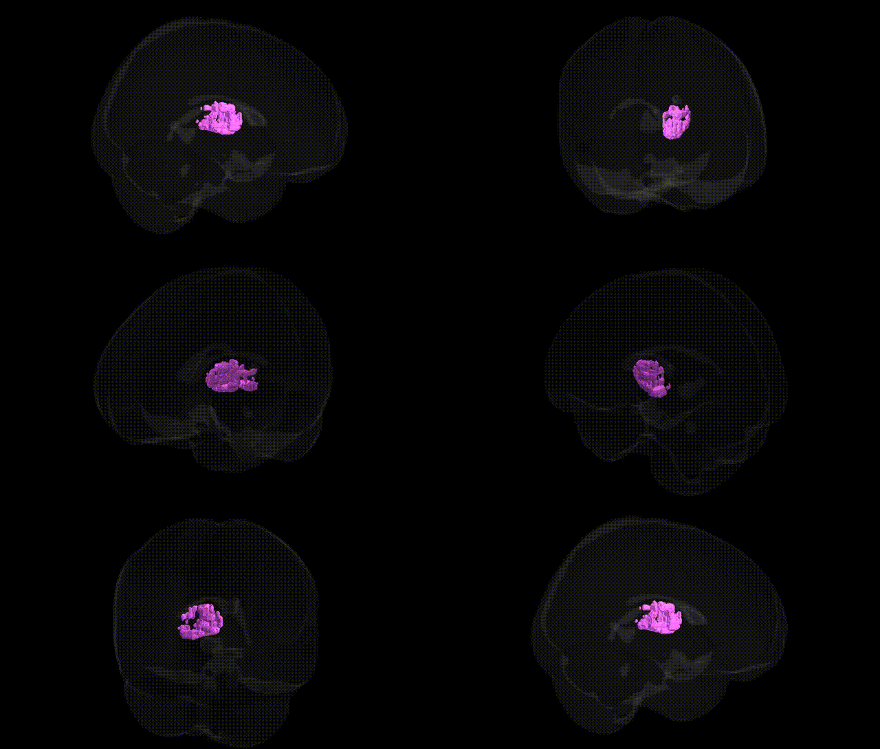
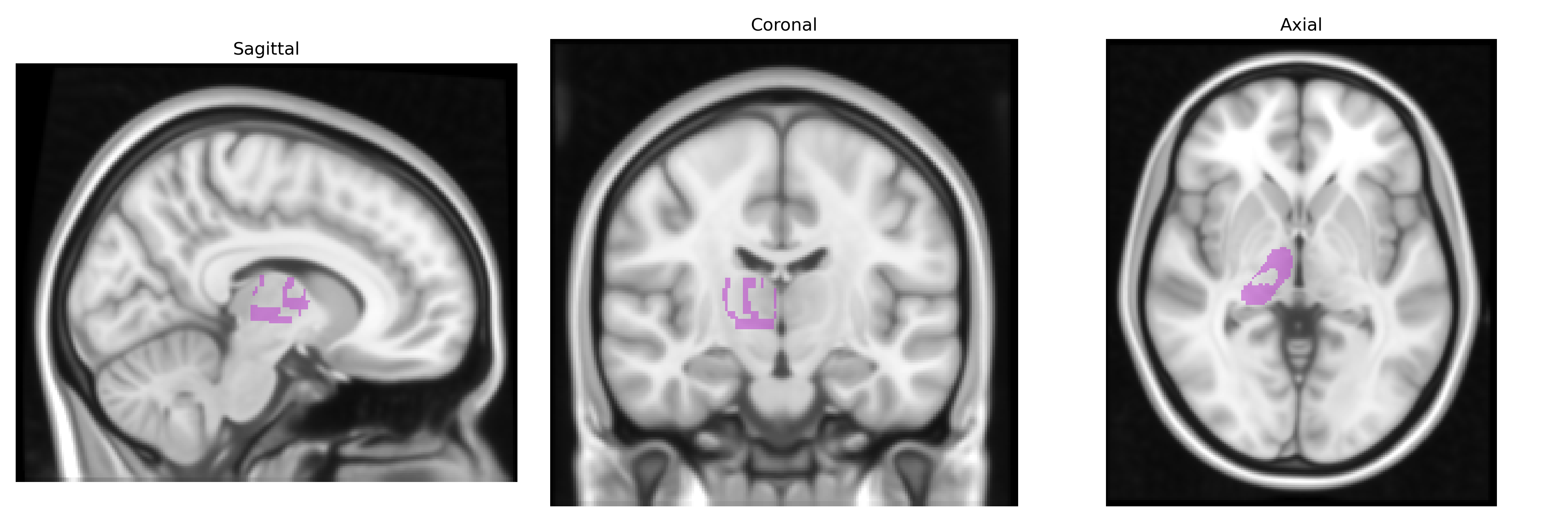
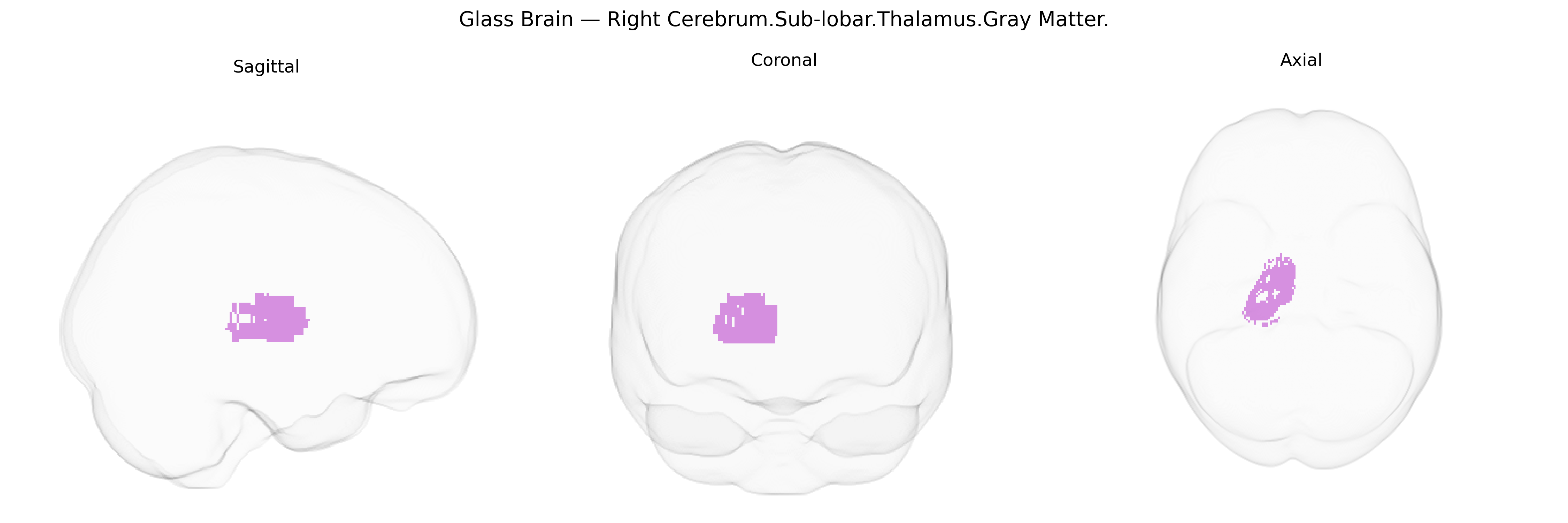
The bilateral Right Cerebrum.Sub-lobar.Thalamus.Gray Matter refers to the gray matter nuclei of the thalamus located within the right cerebral hemisphere’s sub-lobar region, consisting primarily of densely packed neuronal cell bodies that form multiple functionally distinct nuclei. These nuclei serve as critical relay and integration centers for sensory (excluding most olfactory) and motor information, transmitting signals between subcortical structures and widespread cortical areas, including primary sensory cortices, association cortices, and motor regions. The right thalamic gray matter contributes to sensory perception, motor control, attention, arousal, and aspects of cognition and emotion through its extensive reciprocal connections with the cortex, basal ganglia, limbic system, and brainstem. Functionally, it plays a central role in modulating the flow of information to the cortex, synchronizing cortical activity, and participating in network dynamics underlying consciousness and sleep–wake regulation. Thalamus
The bilateral right thalamus (Talairach 1 mm “Right Cerebrum.Sub-lobar.Thalamus.Gray Matter”) shows robust heritability in twin and family studies, and multiple GWAS have identified common variants associated with thalamic volume and shape, particularly in large consortia such as ENIGMA and UK Biobank. Variants in or near genes involved in neurodevelopment, synaptic transmission, and axonal guidance—such as NPTN, GRIN2A, SLC39A8, DLG2, and several loci within MHC and glutamatergic pathways—have been implicated in thalamic structural differences. Polygenic risk scores for schizophrenia, bipolar disorder, and major depressive disorder correlate with altered thalamic volumes or connectivity, and risk loci for schizophrenia (e.g., CACNA1C, MIR137-related regions) and autism spectrum disorder have been linked to thalamocortical circuit abnormalities. GWAS of neuroimaging endophenotypes report that thalamic volume and microstructure share genetic architecture with cognitive performance, educational attainment, and general intelligence, as well as with neurodegenerative and vascular traits, including Alzheimer’s disease risk, small vessel disease markers, and white-matter hyperintensities. Collectively, these findings support the thalamus as a genetically influenced hub whose variation mediates risk and resilience for a wide range of psychiatric, cognitive, and neurological phenotypes, although most associations are bilateral or global rather than specific to the right side alone.
Overview generated by GPT-4o (2026).
Region ID: 531
Hemisphere: bilateral
Atlas: Talairach labels 1mm
Full Quality Version: Download MP4
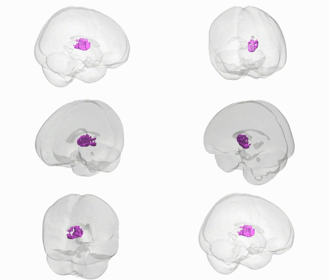
Full Quality Version: Download MP4
Wali Sidiqyar*, Gaurav Rudravaram*, Elyssa M. McMaster, Trent M. Schwartz, Adam M. Saunders, Kurt G. Schilling, Bennett A. Landman "Introducing SPINS: A Shared Public Visualization Library of Neuroanatomical Structures." Medical Imaging with Deep Learning- short paper
This resource is licensed under CC0 1.0 Universal (Public Domain).