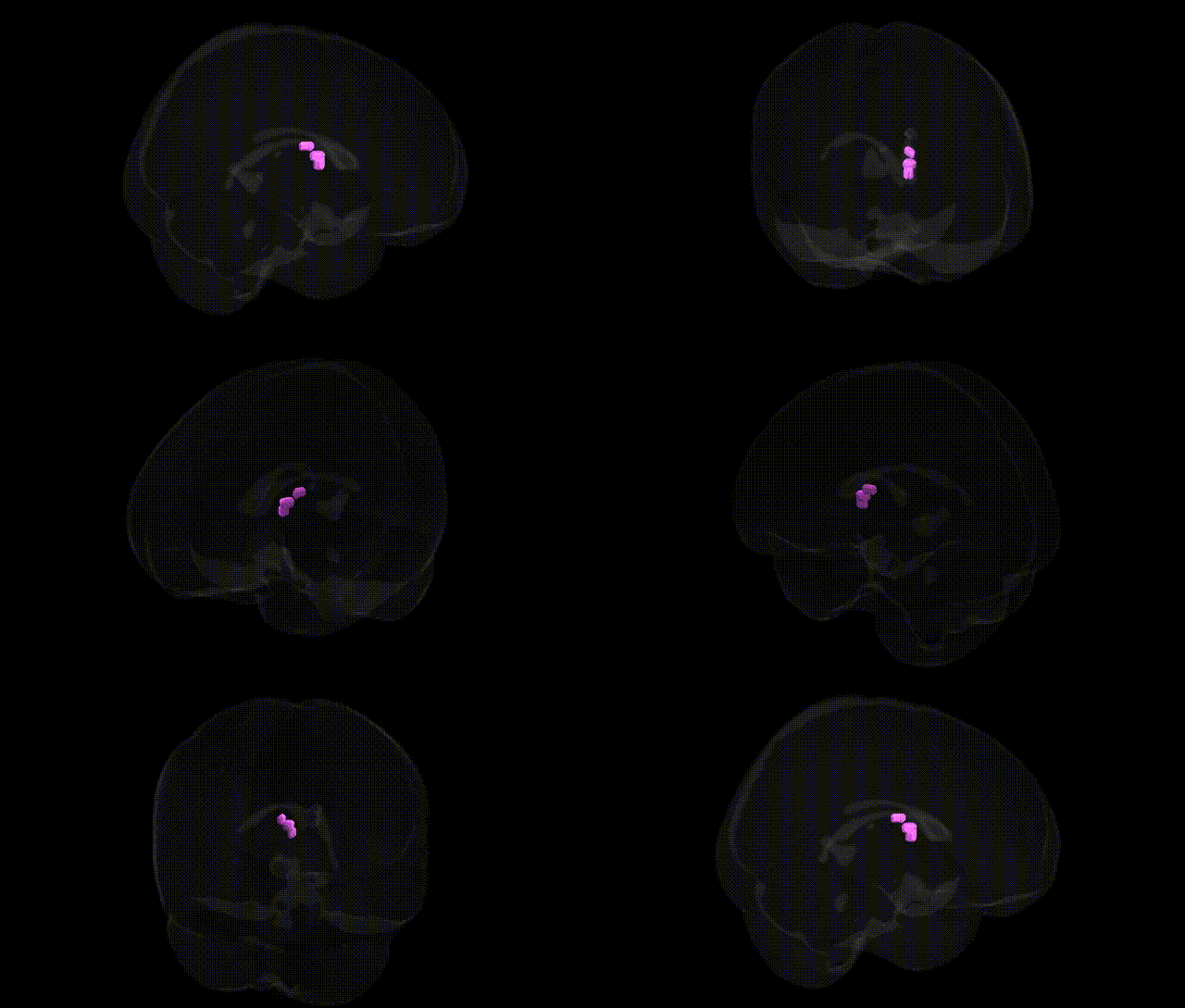
The bilateral Right Cerebrum.Sub-lobar.Thalamus.Gray Matter.Anterior Nucleus corresponds to the anterior nuclear group of the thalamus, a collection of nuclei located in the rostral (anterior) portion of the dorsal thalamus that forms a key node of the limbic system. It receives major input from the mammillary bodies via the mammillothalamic tract and from the hippocampal formation, and projects primarily to the cingulate gyrus and other limbic cortical regions. Functionally, the anterior nucleus is involved in episodic memory, spatial navigation, and emotional processing, and is an integral component of the Papez circuit. Lesions affecting this nucleus are associated with memory impairment and certain forms of diencephalic amnesia, highlighting its role in higher-order cognitive and affective functions within the thalamocortical network. Anterior nuclear group of thalamus
The anterior nucleus of the right thalamus, a key sub-lobar gray matter region, has been implicated in genetic studies primarily through its role in limbic-cortical circuits involved in memory, emotion, and higher-order cognition. GWAS and imaging–genetics work have associated thalamic volume and microstructure with common variants in genes involved in neurodevelopment, synaptic signaling, and axonal guidance (e.g., CACNA1C, CNTNAP2, and several loci near KIAA0319 and GRIN2B), reflecting broader thalamocortical genetic architecture rather than region-unique effects. Polygenic risk scores for schizophrenia, bipolar disorder, major depressive disorder, and attention-deficit/hyperactivity disorder show associations with thalamic structure and connectivity, including anterior thalamic nuclei that relay information to the prefrontal and cingulate cortices, suggesting that risk variants influencing these circuits partly act through thalamic pathways. Thalamic alterations linked to APOE and other Alzheimer’s disease GWAS loci have been reported in the context of memory network degeneration, and variants associated with generalized epilepsy and temporal lobe epilepsy (such as SCN1A and other ion channel genes) have been tied to thalamic involvement in seizure propagation. Additionally, large-scale brain MRI GWAS from consortia like ENIGMA and UK Biobank show that thalamic volumes are moderately heritable and influenced by multiple common variants, some overlapping with loci for cognitive performance, educational attainment, and sleep traits, consistent with the anterior thalamus’s participation in distributed networks governing cognition and arousal rather than a single disease-specific genetic signature.
Overview generated by GPT-4o (2026).
Region ID: 684
Hemisphere: bilateral
Atlas: Talairach labels 1mm
Full Quality Version: Download MP4
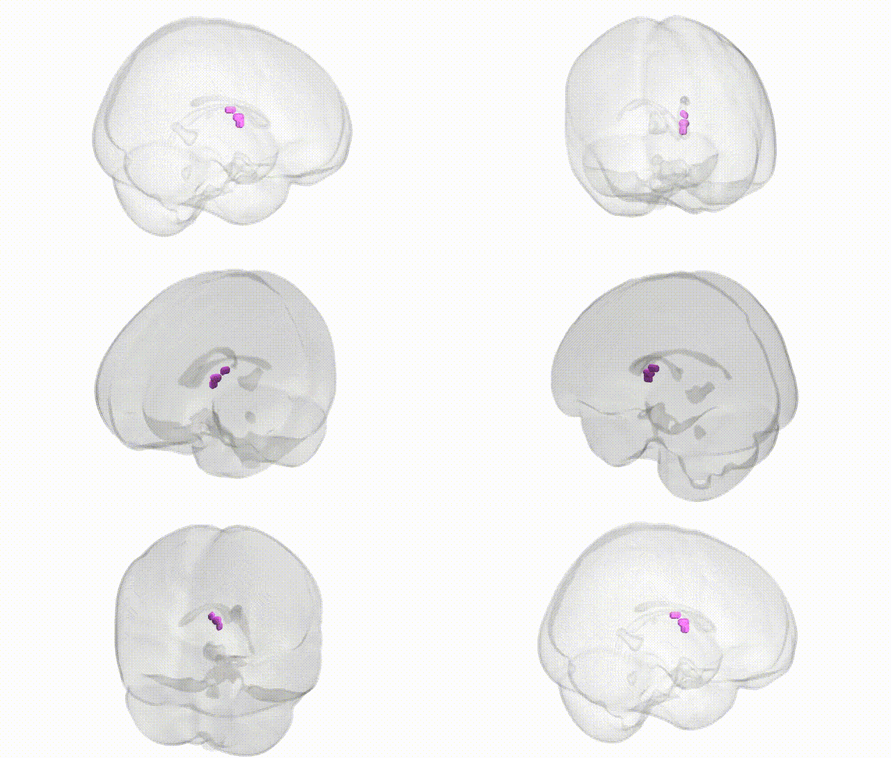
Full Quality Version: Download MP4
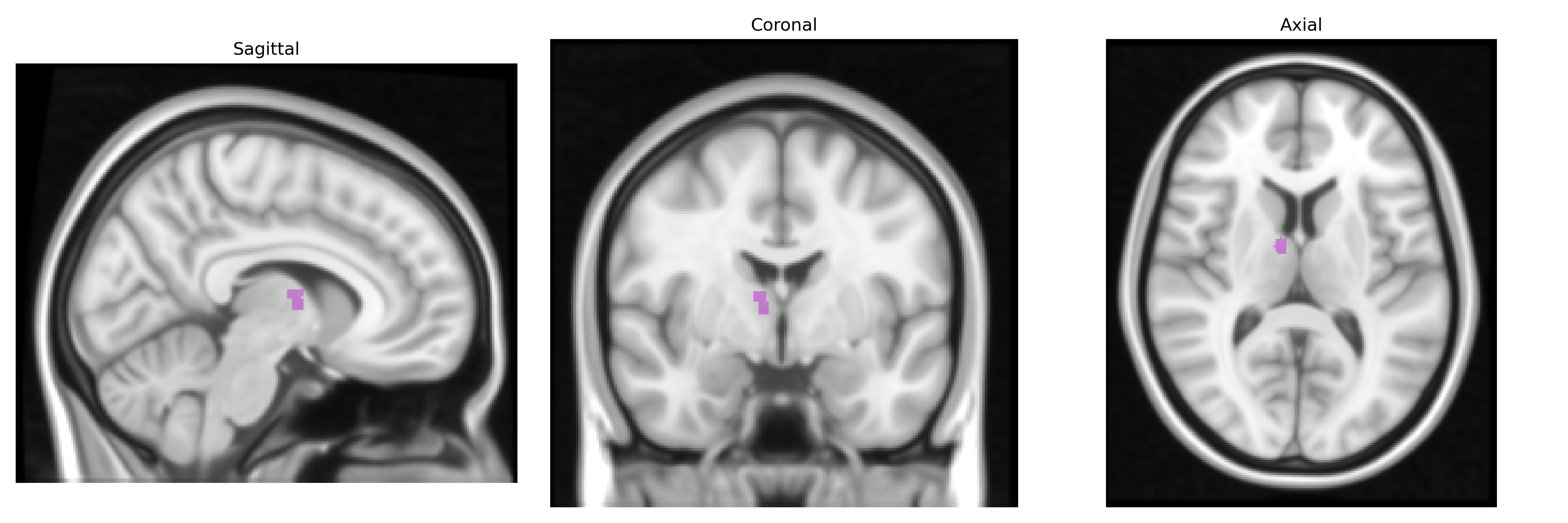
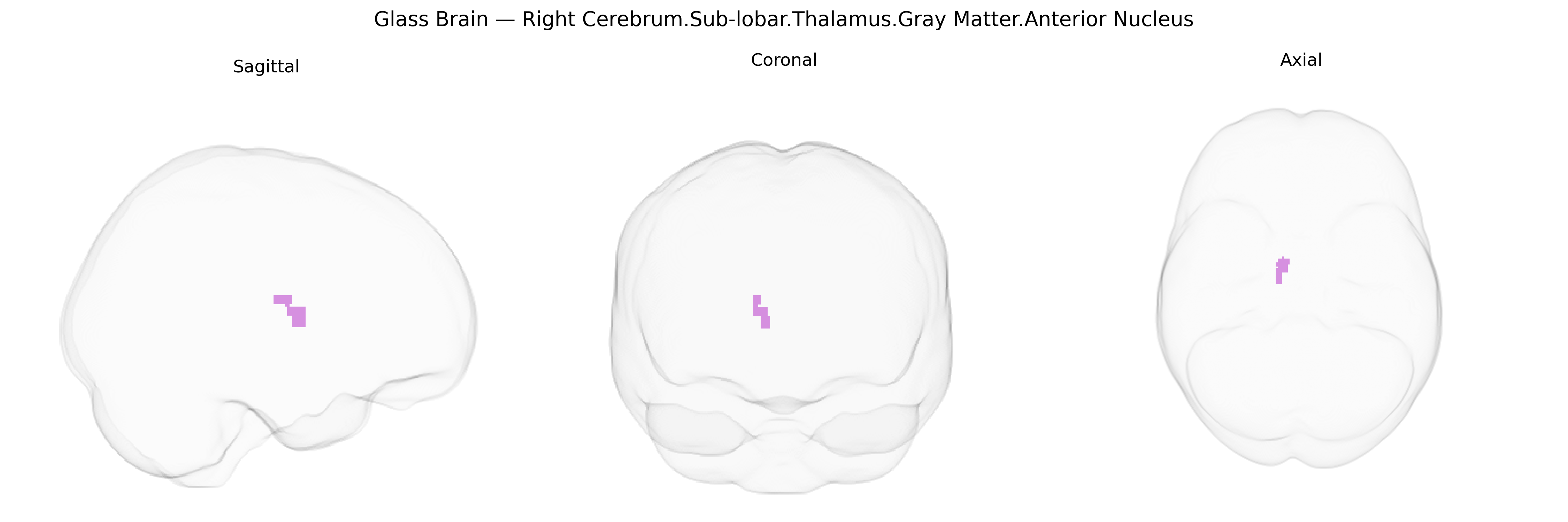
Wali Sidiqyar*, Gaurav Rudravaram*, Elyssa M. McMaster, Trent M. Schwartz, Adam M. Saunders, Kurt G. Schilling, Bennett A. Landman "Introducing SPINS: A Shared Public Visualization Library of Neuroanatomical Structures." Medical Imaging with Deep Learning- short paper
This resource is licensed under CC0 1.0 Universal (Public Domain).