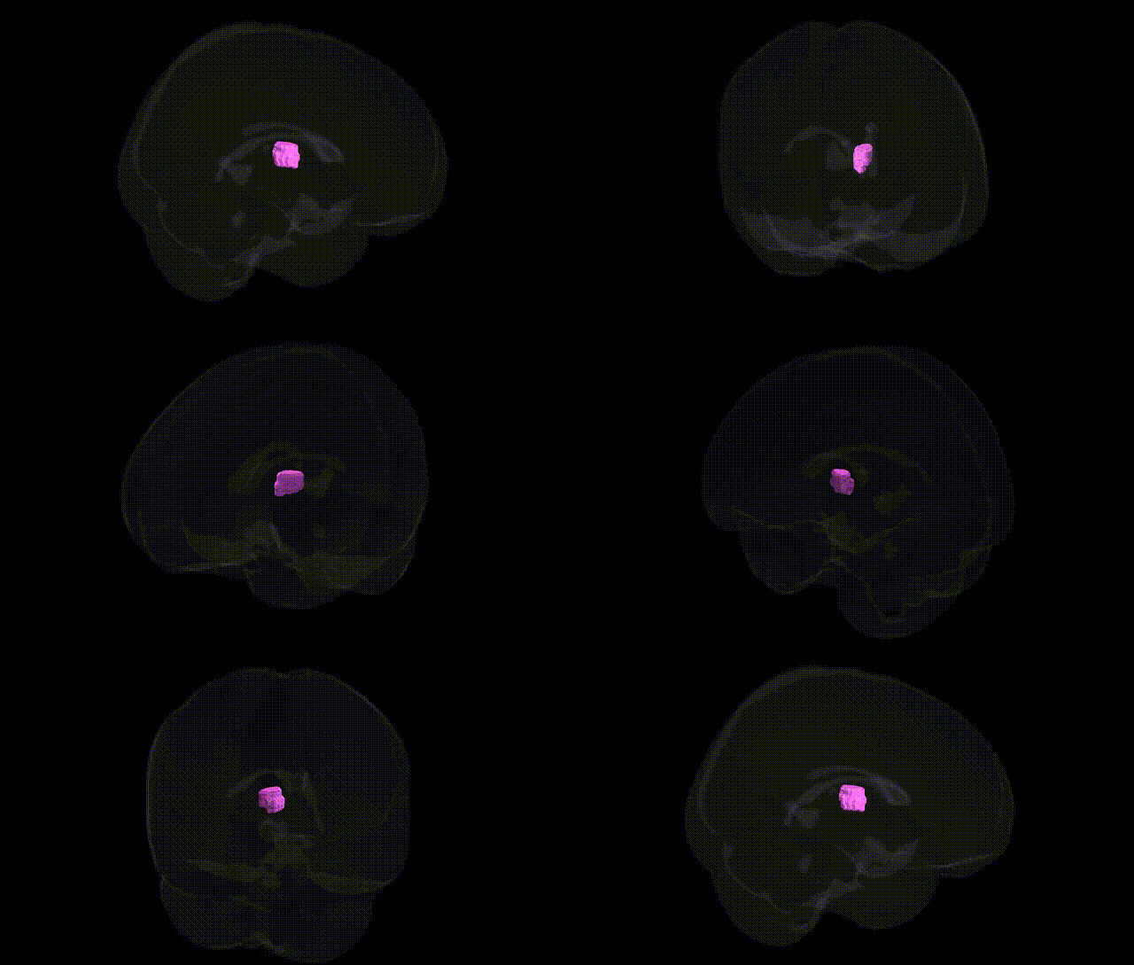
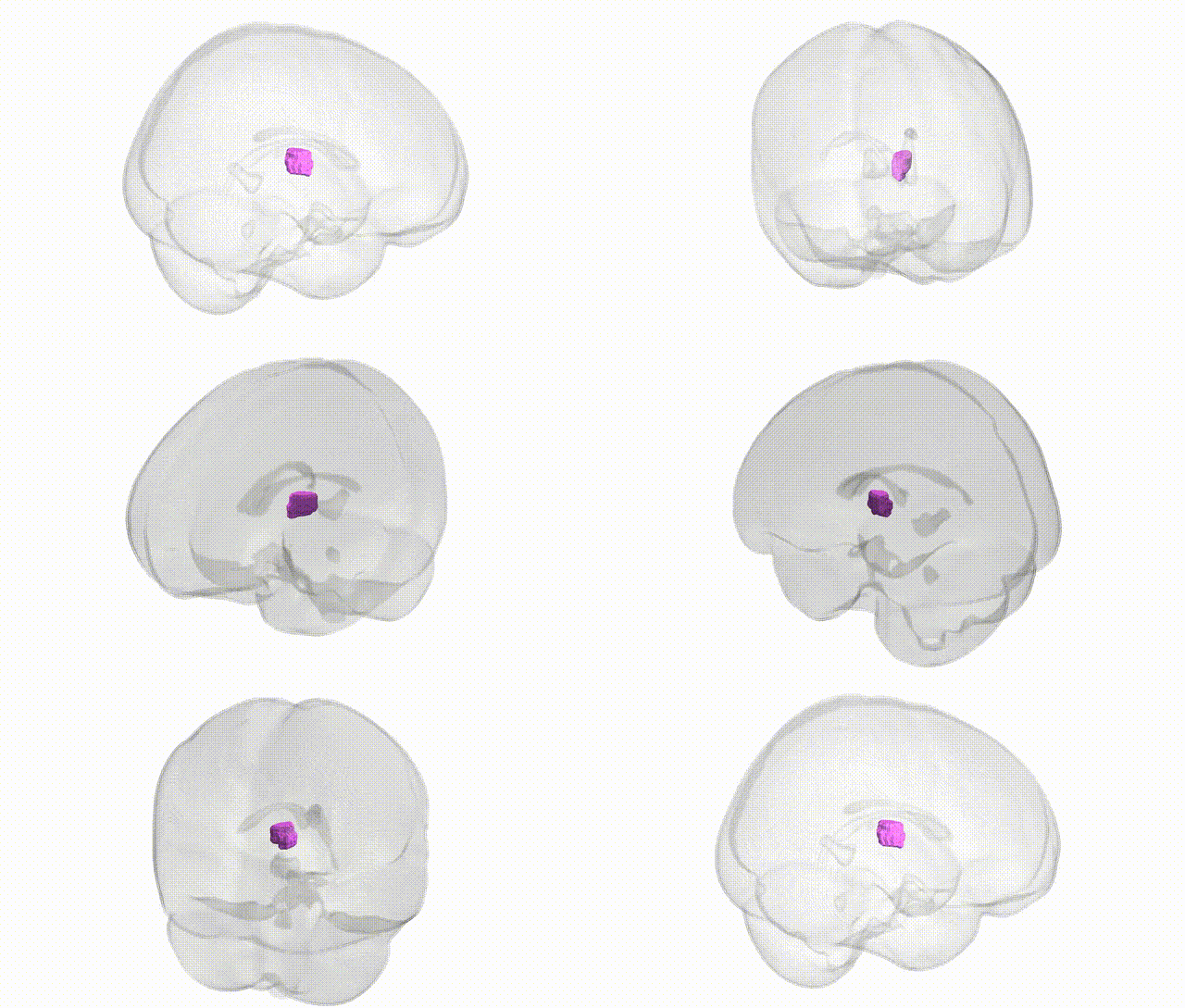
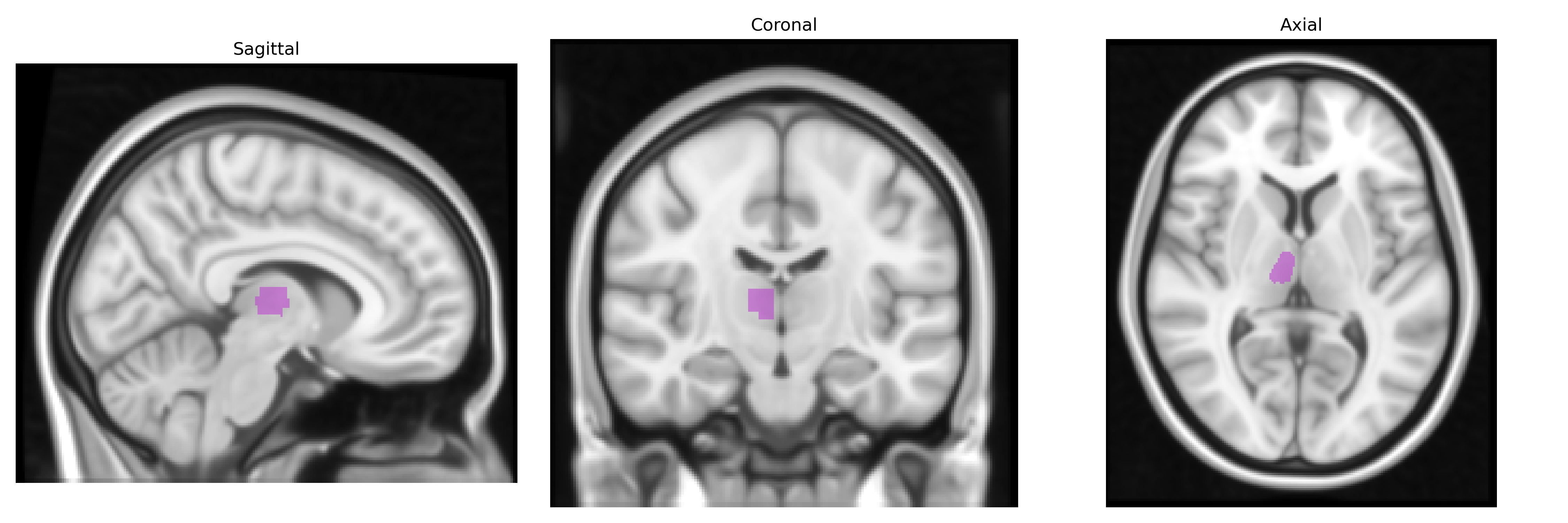
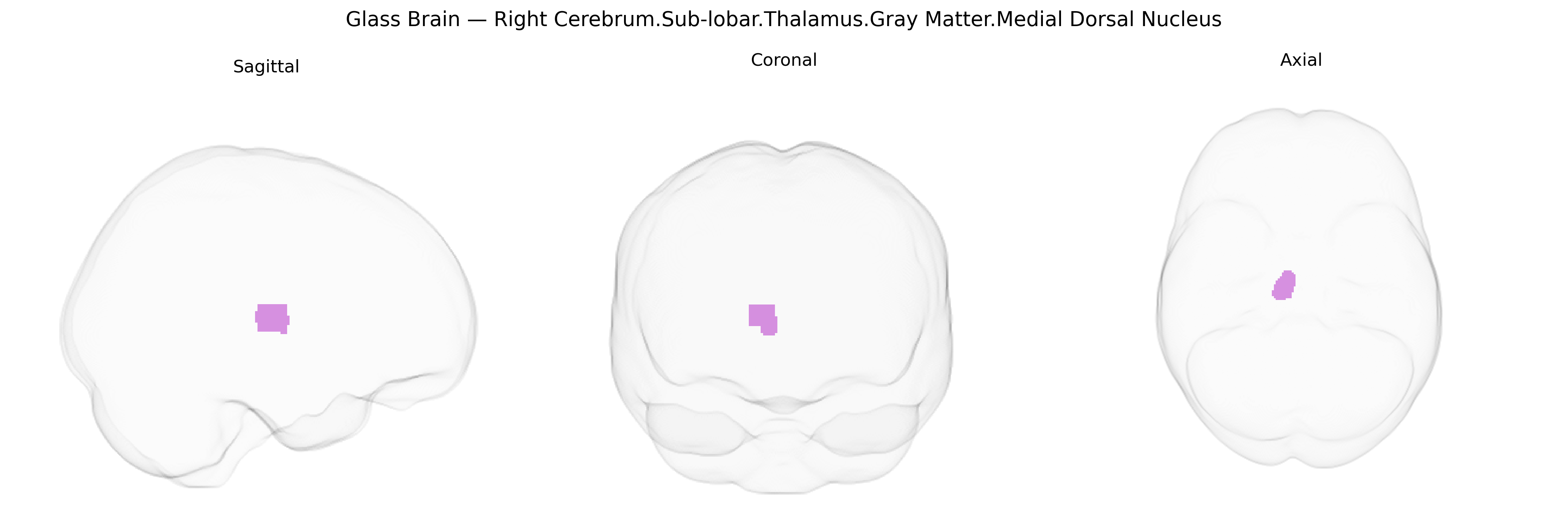
The bilateral Right Cerebrum.Sub-lobar.Thalamus.Gray Matter.Medial Dorsal Nucleus corresponds to the mediodorsal (medial dorsal) nucleus of the thalamus, a higher-order relay nucleus situated in the dorsal thalamus that is heavily interconnected with prefrontal, cingulate, and limbic cortices. It receives inputs from basal ganglia (particularly the globus pallidus and substantia nigra), amygdala, and midbrain structures, and projects extensively to the prefrontal cortex, thereby contributing to executive functions, working memory, decision-making, and emotional processing. The mediodorsal nucleus is critically involved in integrating cognitive and affective information and is implicated in neuropsychiatric conditions such as schizophrenia, mood disorders, and memory impairments, where disruption of thalamo-prefrontal circuitry is observed. Mediodorsal nucleus
Genetic associations involving the medial dorsal (mediodorsal) thalamic nucleus—a key subcortical node for cognition, affect, and fronto-thalamic circuitry—have emerged largely from imaging-genetics and GWAS of subcortical volumes and neuropsychiatric traits. Large ENIGMA and UK Biobank–based GWAS have identified common variants affecting thalamic (including medial dorsal) volume and connectivity, implicating loci near or within genes such as HMGA2, FAT3, DCC, and several calcium channel and synaptic genes, though regional specificity to the medial dorsal nucleus is often inferred from parcellation-based analyses rather than directly labeled. Polygenic overlap links thalamic structure with schizophrenia, bipolar disorder, major depressive disorder, ADHD, and autism spectrum disorder, consistent with repeated reports of altered medial dorsal volume, microstructure, or functional connectivity in these conditions. Genetic liability for cognitive traits (e.g., intelligence, working memory, educational attainment) shows correlated effects on thalamic nuclei volumes and fronto-thalamic network integrity, suggesting that variants influencing neuronal development, axon guidance, and synaptic plasticity contribute to interindividual differences in medial dorsal thalamic anatomy. Additional GWAS have related thalamic structure and connectivity—including medial dorsal components—to risk loci for Parkinson’s and essential tremor, as well as sleep and circadian traits, underscoring this nucleus’s role as a genetically modulated hub linking cortical, limbic, and motor systems.
Overview generated by GPT-4o (2026).
Region ID: 637
Hemisphere: bilateral
Atlas: Talairach labels 1mm
Full Quality Version: Download MP4
Full Quality Version: Download MP4
Wali Sidiqyar*, Gaurav Rudravaram*, Elyssa M. McMaster, Trent M. Schwartz, Adam M. Saunders, Kurt G. Schilling, Bennett A. Landman "Introducing SPINS: A Shared Public Visualization Library of Neuroanatomical Structures." Medical Imaging with Deep Learning- short paper
This resource is licensed under CC0 1.0 Universal (Public Domain).