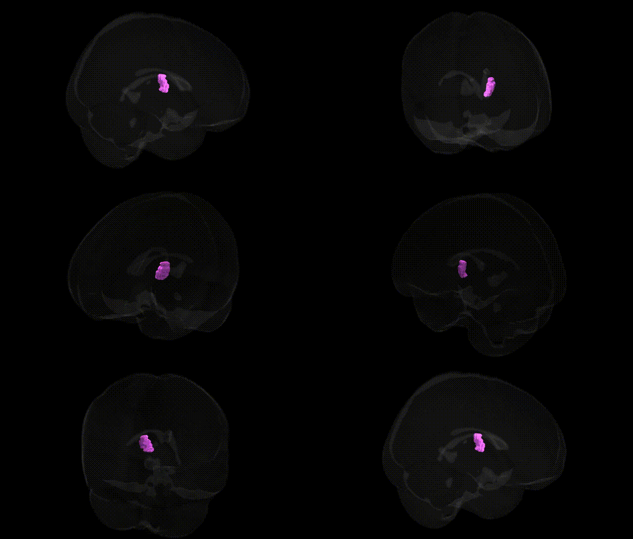
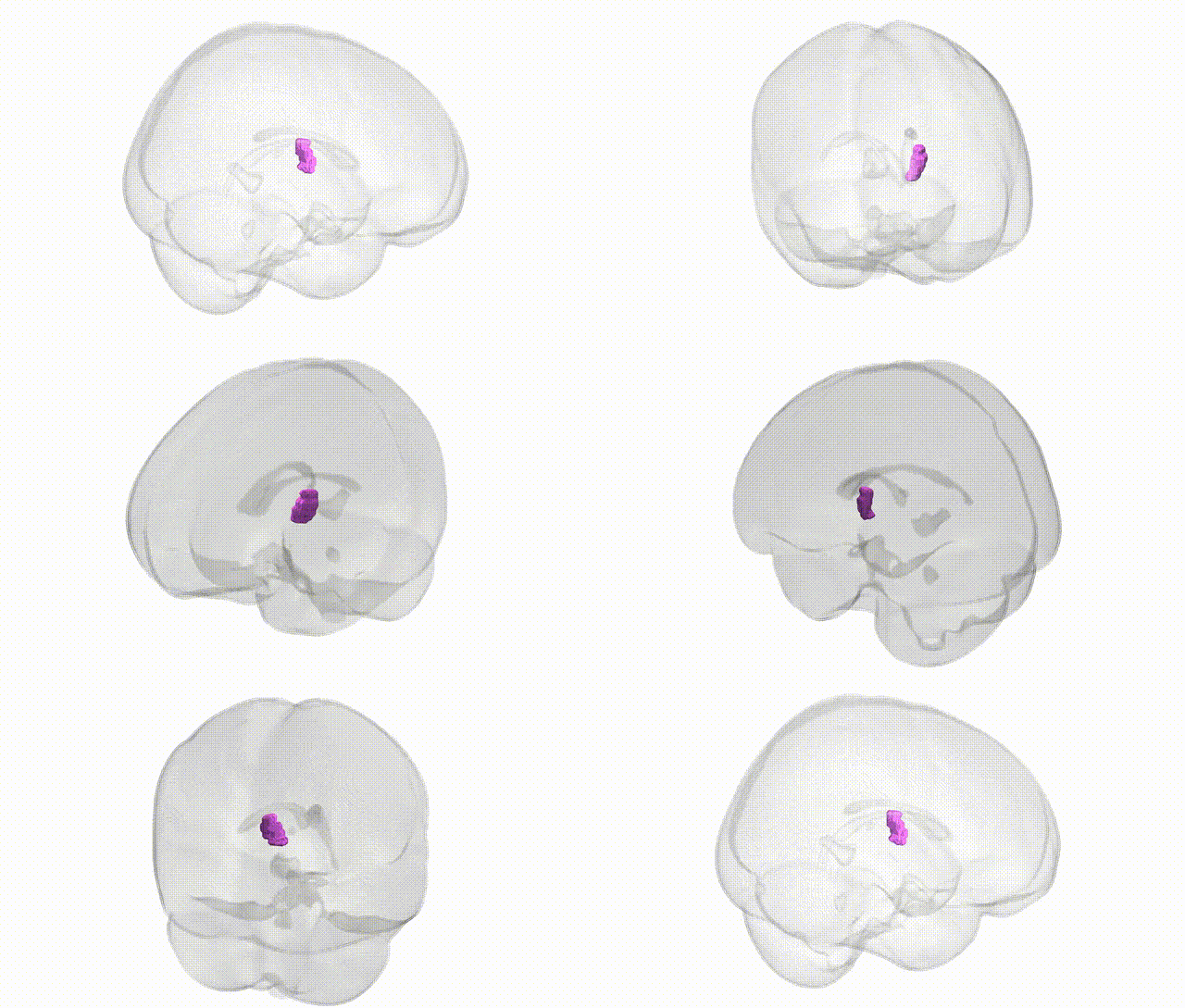
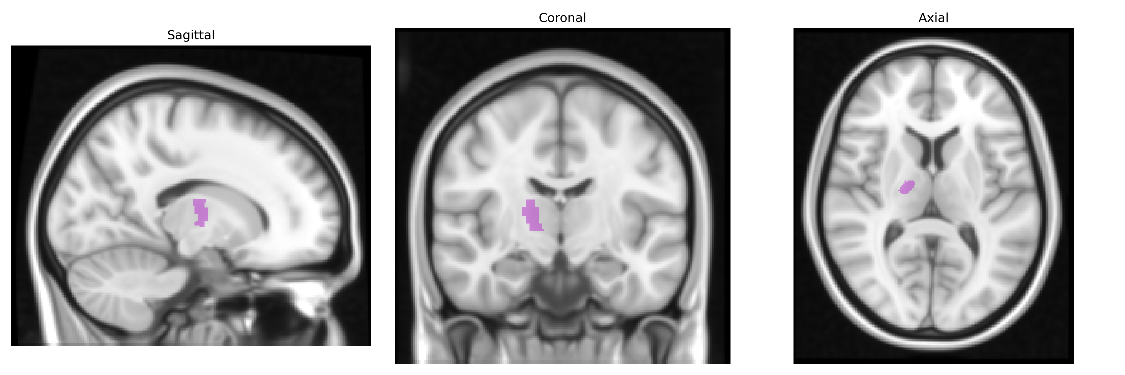
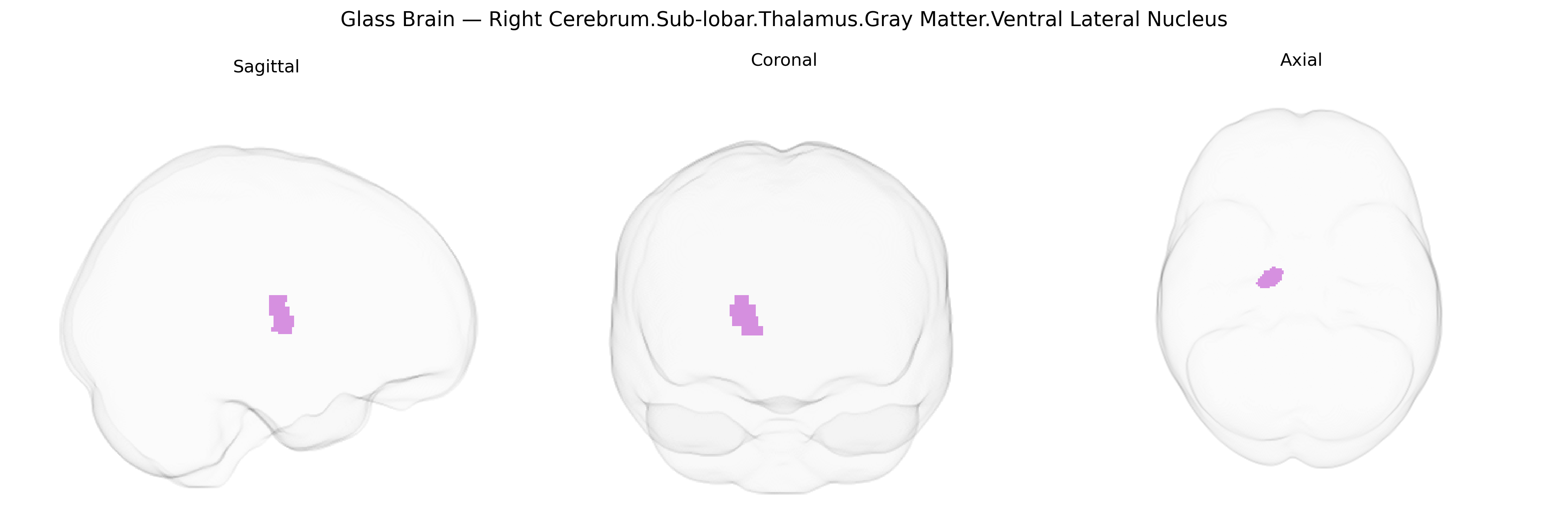
The bilateral Right Cerebrum.Sub-lobar.Thalamus.Gray Matter.Ventral Lateral Nucleus corresponds to the ventral lateral (VL) nucleus of the thalamus, a major motor relay hub situated in the subcortical gray matter of the diencephalon. It receives prominent input from the cerebellum and globus pallidus and projects primarily to motor and premotor cortices, thereby integrating cerebellar and basal ganglia signals to modulate movement planning, execution, and coordination. Functionally, the VL nucleus participates in the regulation of voluntary motor activity, muscle tone, and fine motor control, and is often implicated in movement disorders and targeted in certain neurosurgical interventions for tremor. This region forms part of the larger thalamic motor circuit and contributes to sensorimotor integration through its extensive reciprocal connections with cortical and subcortical motor areas. Ventral lateral nucleus of thalamus
The ventral lateral (VL) nucleus of the thalamus, as defined in the Talairach 1 mm atlas within the right sub-lobar gray matter, has been implicated indirectly in several genetic and GWAS-based associations through its roles in motor, premotor, and cerebellar-cortical circuitry rather than through VL-specific locus tagging. Variants in genes influencing thalamocortical development and excitability (e.g., CACNA1A, CACNA1H, GRIN2B, GAD1) and broader synaptic or neurodevelopmental pathways (e.g., CNTNAP2, DISC1, BDNF) have been associated with disorders in which VL-thalamic dysfunction and altered thalamocortical connectivity play a key role, including generalized and focal epilepsies, essential tremor, Parkinson’s disease, dystonia, Tourette syndrome, schizophrenia, bipolar disorder, major depression, and ADHD. GWAS of intracranial or subcortical volumes and thalamic morphology (such as ENIGMA and UK Biobank–based studies) have identified polygenic influences on thalamic size and microstructure (involving loci near genes like NPTN, FAM101B, and others involved in axon guidance, myelination, and synaptic function), though these analyses typically treat the thalamus as a whole or use diffusion-based thalamic nuclei parcellations rather than specifically isolating the VL nucleus. Imaging–genetics work combining fMRI or structural MRI with polygenic risk scores for schizophrenia, depression, or movement disorders has linked higher polygenic burden to altered thalamic connectivity and volume, including in motor and premotor thalamic territories that overlap the VL region. Overall, genetic evidence currently supports a polygenic, system-level contribution to VL-thalamic structure and function through genes regulating ion channels, synaptic transmission, neurodevelopment, and cortical–subcortical connectivity, with associations emerging primarily via broader thalamic or motor-network GWAS rather than nucleus-specific genetic mapping.
Overview generated by GPT-4o (2026).
Region ID: 644
Hemisphere: bilateral
Atlas: Talairach labels 1mm
Full Quality Version: Download MP4
Full Quality Version: Download MP4
Wali Sidiqyar*, Gaurav Rudravaram*, Elyssa M. McMaster, Trent M. Schwartz, Adam M. Saunders, Kurt G. Schilling, Bennett A. Landman "Introducing SPINS: A Shared Public Visualization Library of Neuroanatomical Structures." Medical Imaging with Deep Learning- short paper
This resource is licensed under CC0 1.0 Universal (Public Domain).