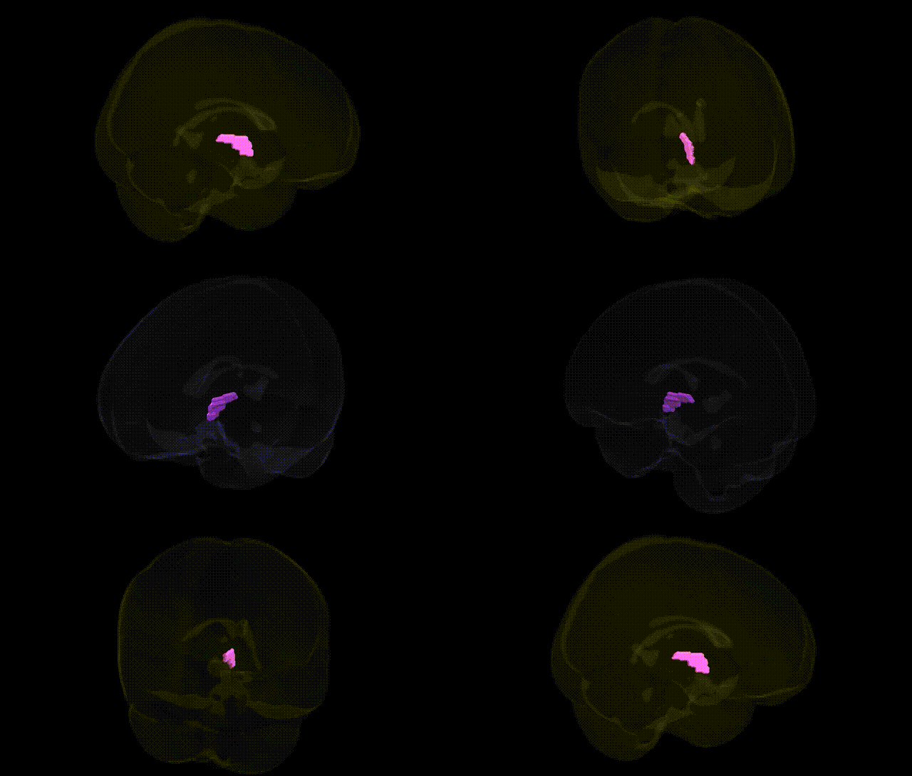
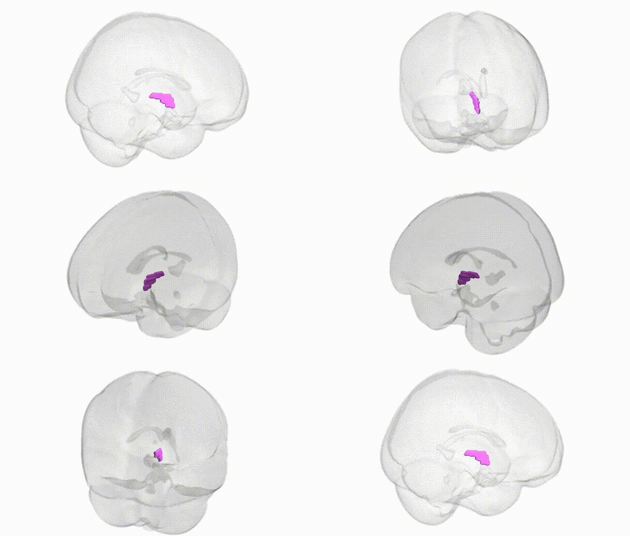
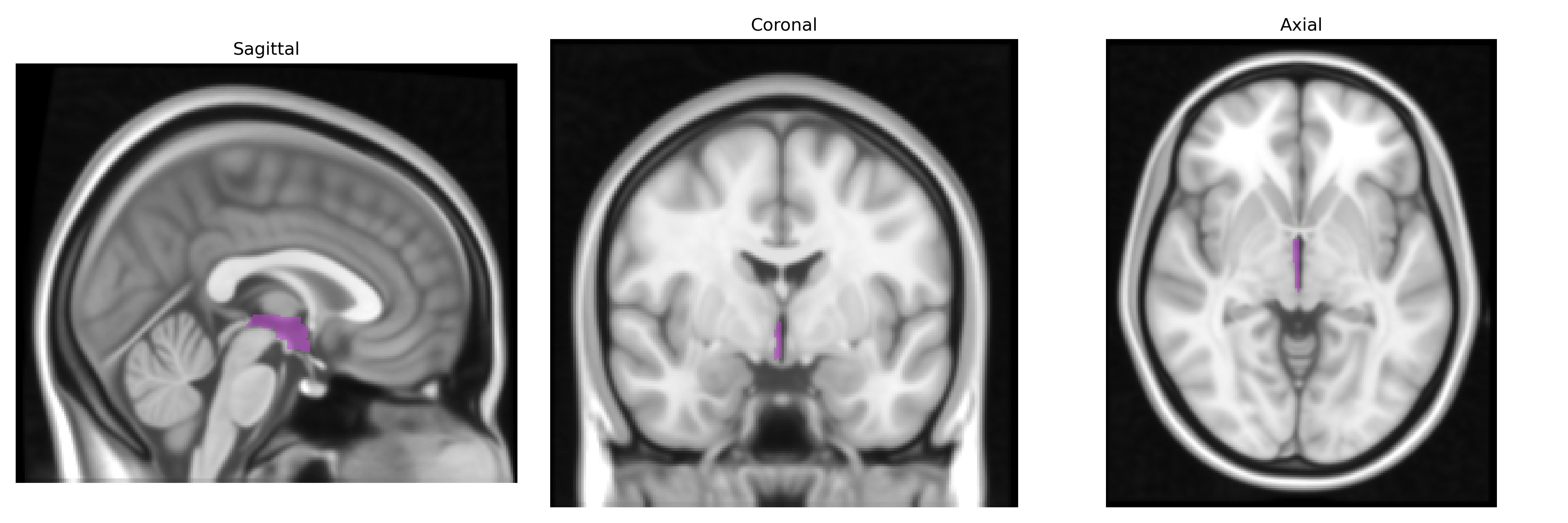
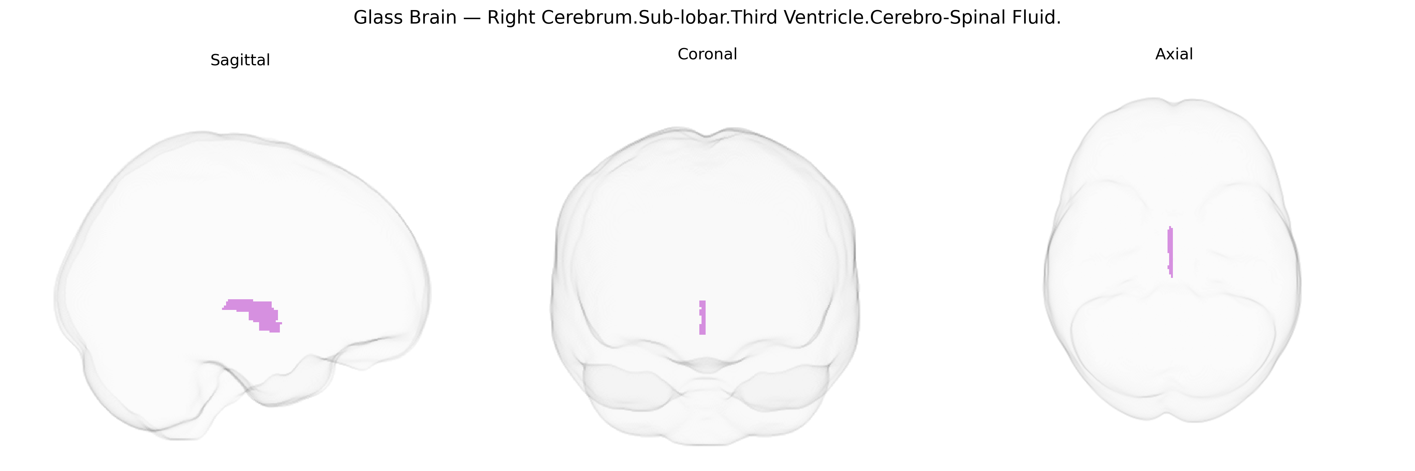
The bilateral Right Cerebrum.Sub-lobar.Third Ventricle.Cerebro-Spinal Fluid region corresponds to cerebrospinal fluid (CSF) occupying the third ventricle, a narrow midline cavity located between the two halves of the diencephalon, bordered primarily by the thalamus and hypothalamus. In the Talairach 1 mm atlas, this label captures voxels containing CSF rather than neural parenchyma, reflecting the internal ventricular space through which CSF circulates from the lateral ventricles to the cerebral aqueduct and subsequently to the fourth ventricle. Functionally, the third ventricle and its CSF play roles in cushioning the brain, maintaining intracranial pressure, facilitating waste clearance, and serving as a conduit for neuroactive substances and metabolites within the ventricular system. There is no direct link for this composite label; a related structure is the Third ventricle.
The bilateral Right Cerebrum.Sub-lobar.Third Ventricle.Cerebro-Spinal Fluid region in the Talairach 1 mm atlas corresponds anatomically to the cerebrospinal fluid–filled third ventricle and its immediate periventricular surroundings rather than gray matter nuclei, so genetic associations typically arise from GWAS of ventricular or CSF volumes rather than this label per se. Large imaging–genetics studies (e.g., ENIGMA, UK Biobank) have identified multiple loci influencing lateral and third ventricular size, including variants in or near genes related to neurodevelopment and brain morphogenesis (such as PLEKHG1, TRIM47, MAPT, and other loci in 17q21, as well as genes affecting cilia function and CSF dynamics), although findings are less consistently specific to the third ventricle alone. Increased ventricular/CSF volume—often interpreted as a proxy for global or regional brain atrophy—has been genetically correlated with polygenic risk for neurodegenerative and psychiatric conditions, including Alzheimer’s disease, schizophrenia, and bipolar disorder, and with common variants in APOE and other Alzheimer’s risk loci that show parallel effects on medial temporal and global atrophy. Ventricular and CSF volume heritability is moderate to high, and GWAS indicate shared genetic architectures with intracranial volume, cortical thickness, and subcortical structures; however, most studies report global or lateral ventricular measures, so specific, reproducible gene–third ventricle associations remain relatively coarse and largely reflect broader neurodevelopmental and neurodegenerative genetic influences rather than a distinct, uniquely third-ventricle–restricted genetic signature.
Overview generated by GPT-4o (2026).
Region ID: 360
Hemisphere: bilateral
Atlas: Talairach labels 1mm
Full Quality Version: Download MP4
Full Quality Version: Download MP4
Wali Sidiqyar*, Gaurav Rudravaram*, Elyssa M. McMaster, Trent M. Schwartz, Adam M. Saunders, Kurt G. Schilling, Bennett A. Landman "Introducing SPINS: A Shared Public Visualization Library of Neuroanatomical Structures." Medical Imaging with Deep Learning- short paper
This resource is licensed under CC0 1.0 Universal (Public Domain).