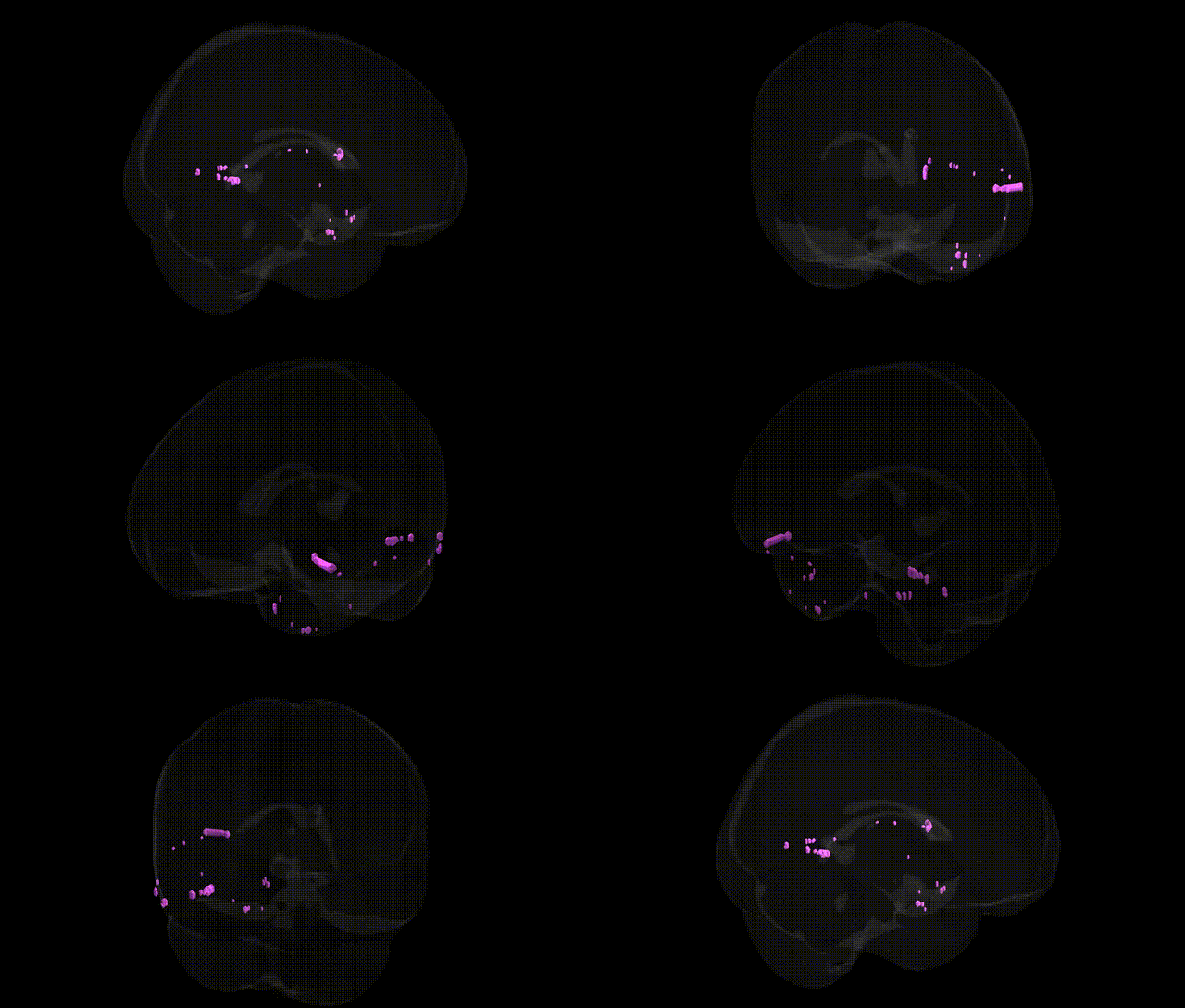
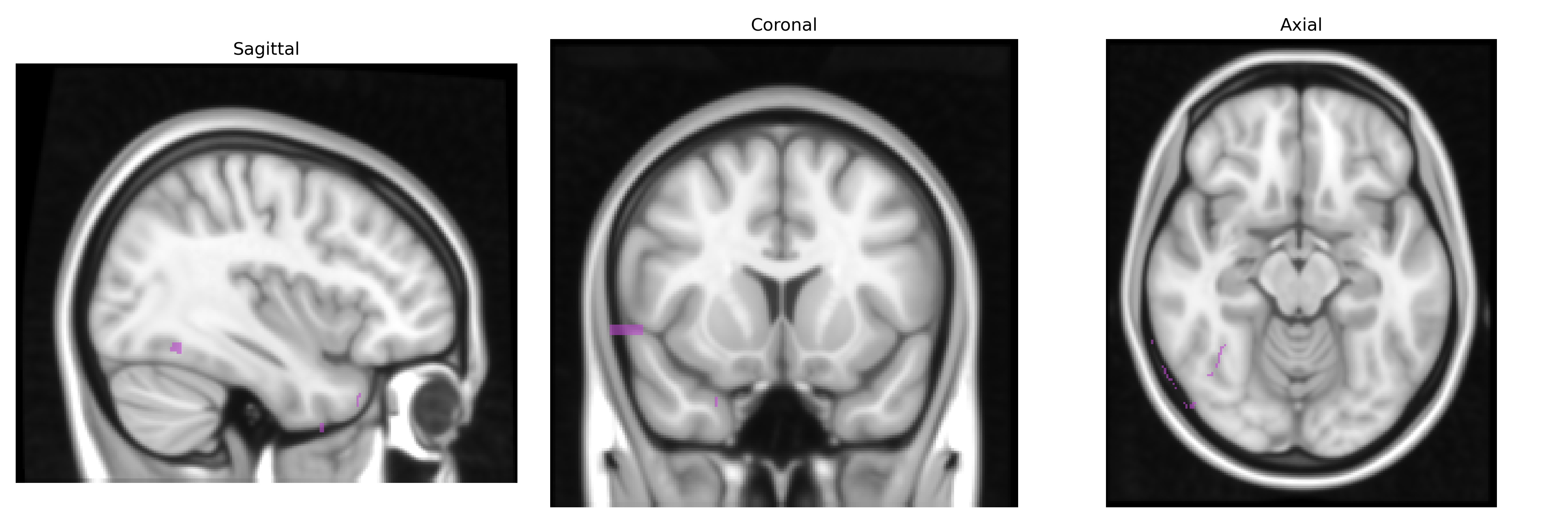
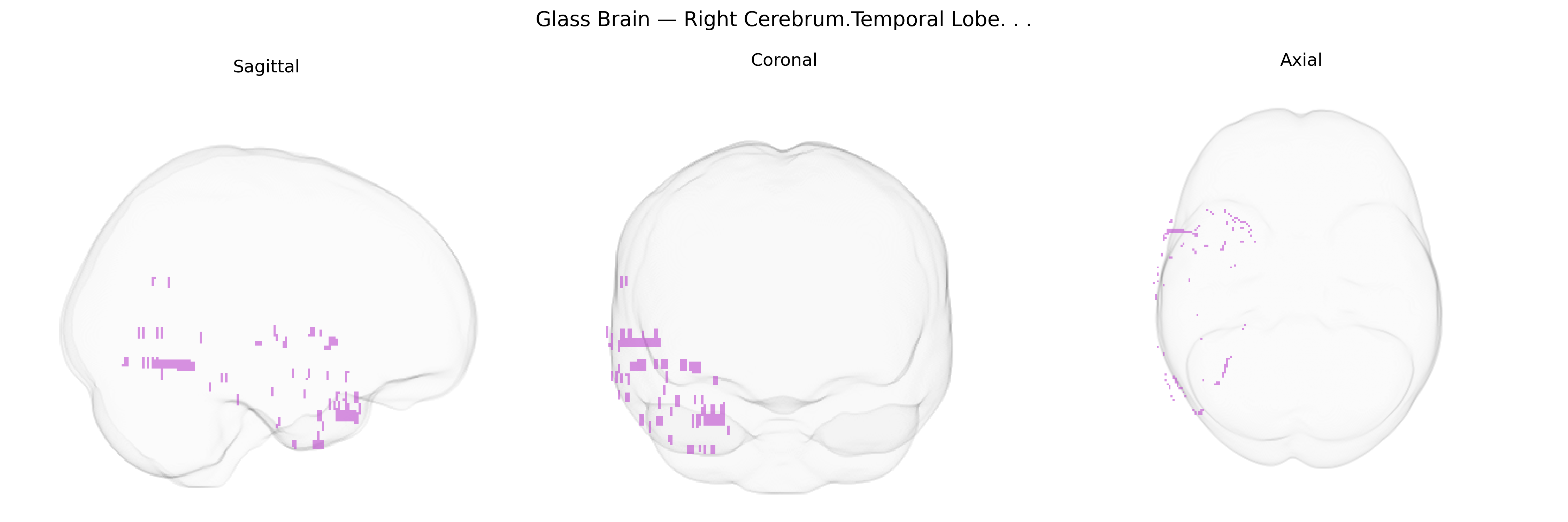
The bilateral Right Cerebrum.Temporal Lobe corresponds to cortical and subcortical territories in the right temporal lobe, including portions of the superior, middle, and inferior temporal gyri, as well as deeper structures such as parts of the hippocampal formation and amygdaloid complex, depending on the exact Talairach coordinate. Functionally, this region is involved in high-level auditory processing, aspects of language (particularly prosody and comprehension of non-literal content), semantic memory, and the integration of multimodal sensory inputs relevant for object and face recognition. The right temporal lobe also contributes to social cognition, emotional processing, and certain forms of episodic memory, with lesions often producing deficits in nonverbal memory, music perception, and aspects of affective prosody. A related overview of this territory is provided in the article on the Temporal lobe.
The bilateral right temporal lobe (right cerebrum, temporal lobe in the Talairach 1 mm atlas) has been implicated in numerous genetic and genome-wide association studies linking its structure, function, and connectivity to diverse traits and disorders. Variants in genes involved in synaptic plasticity and neurodevelopment (for example, BDNF, NRG1, GRIN2B, and CNTNAP2) have been associated with right temporal volume or cortical thickness, while large-scale imaging–genetics consortia (such as ENIGMA and UK Biobank GWAS) have identified multiple loci—often near genes related to axon guidance, cell adhesion, and neurogenesis—that influence temporal lobe morphology. Right temporal structural and functional alterations linked to genetic risk are consistently observed in schizophrenia and bipolar disorder (with contributions from common variants in genes such as CACNA1C, ZNF804A, and MIR137-associated loci), in autism spectrum disorder (including rare CNVs and mutations in synaptic genes like SHANK3 and NRXN1), and in major depressive disorder and anxiety-related traits. Genetic influences on language lateralization and social cognition, including variants in FOXP2-related networks and polygenic scores for educational attainment and cognitive ability, have been associated with asymmetries and connectivity patterns involving the right temporal cortex, while Alzheimer’s disease and other dementias show GWAS-supported links (e.g., APOE and additional loci) to right temporal atrophy and tau/amyloid-related neurodegeneration affecting this region.
Overview generated by GPT-4o (2026).
Region ID: 11
Hemisphere: bilateral
Atlas: Talairach labels 1mm
Full Quality Version: Download MP4
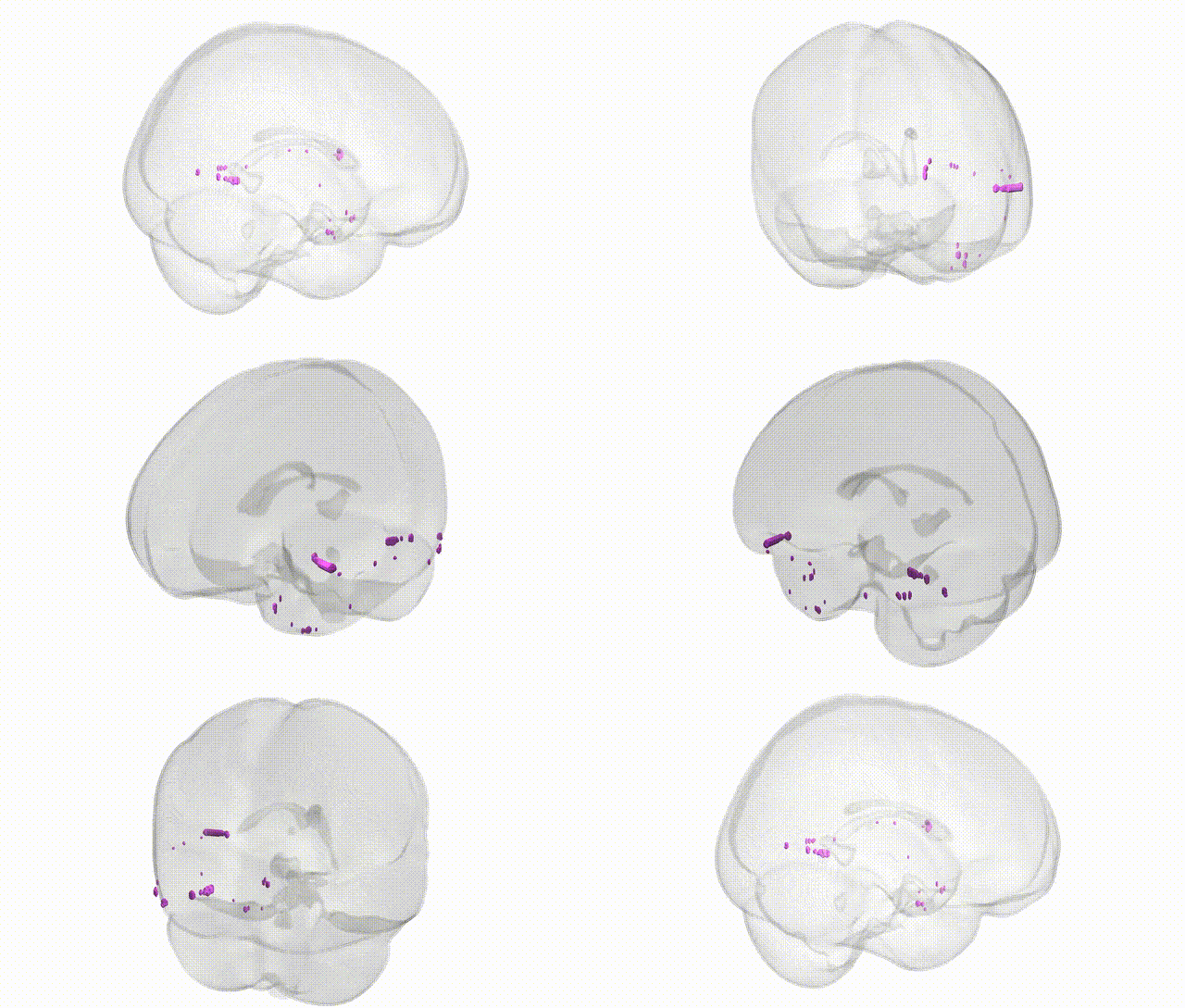
Full Quality Version: Download MP4
Wali Sidiqyar*, Gaurav Rudravaram*, Elyssa M. McMaster, Trent M. Schwartz, Adam M. Saunders, Kurt G. Schilling, Bennett A. Landman "Introducing SPINS: A Shared Public Visualization Library of Neuroanatomical Structures." Medical Imaging with Deep Learning- short paper
This resource is licensed under CC0 1.0 Universal (Public Domain).