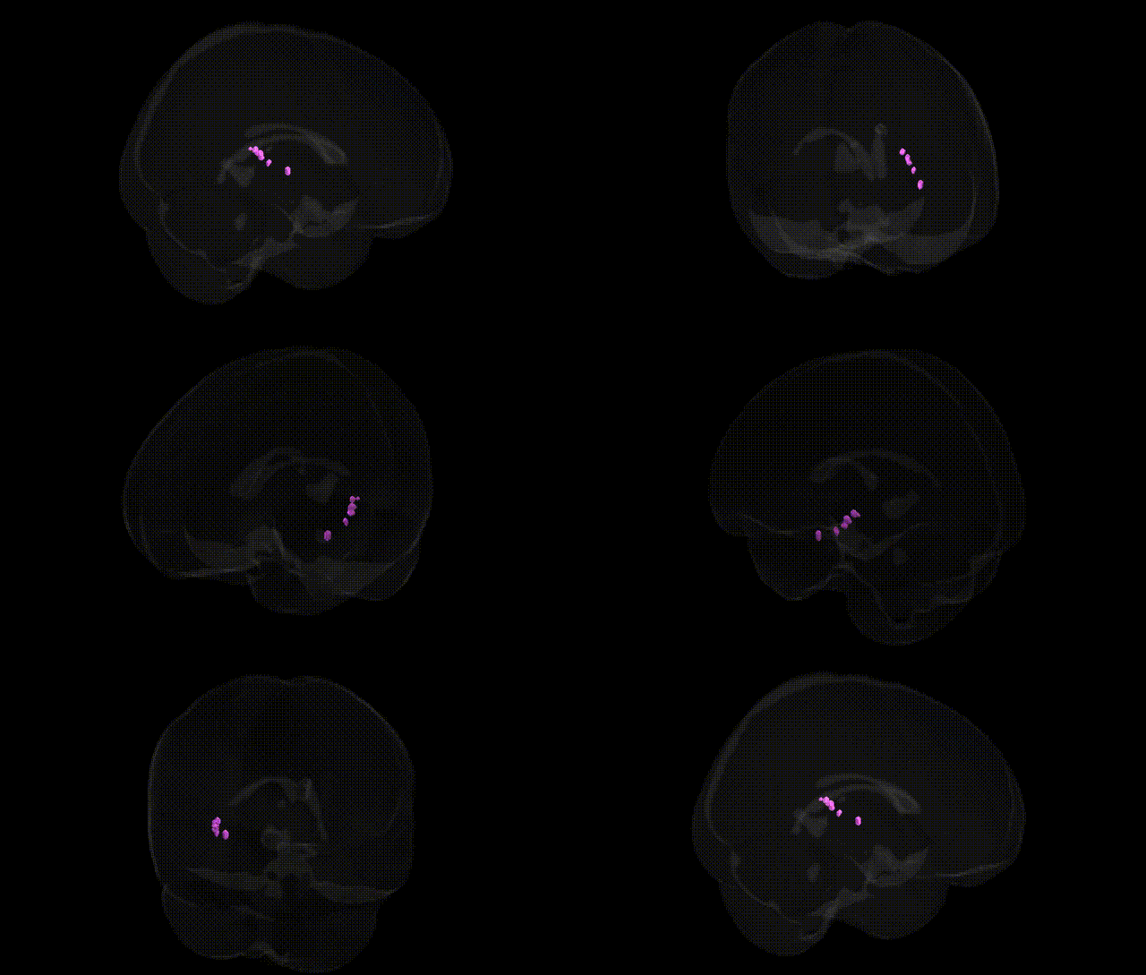
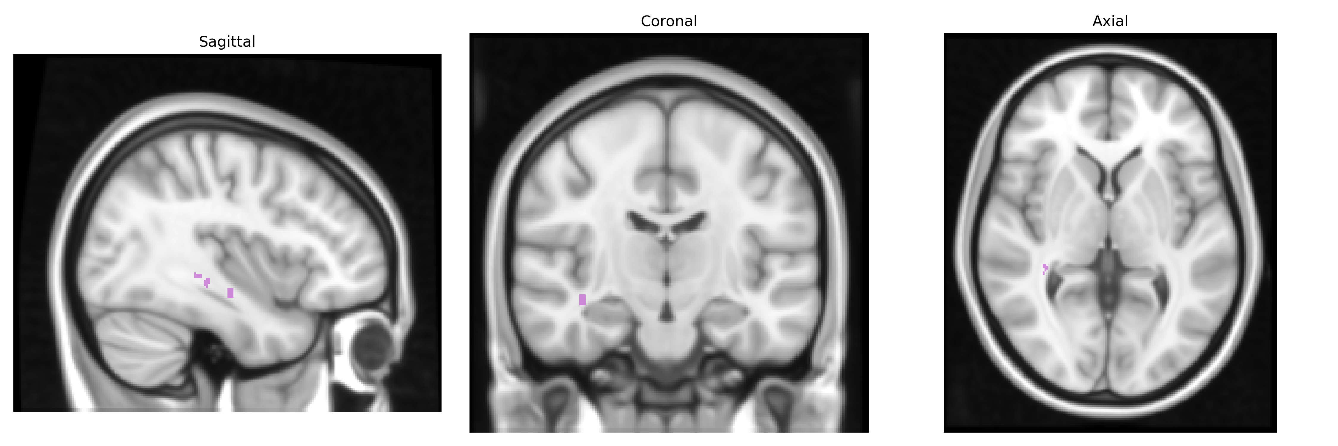
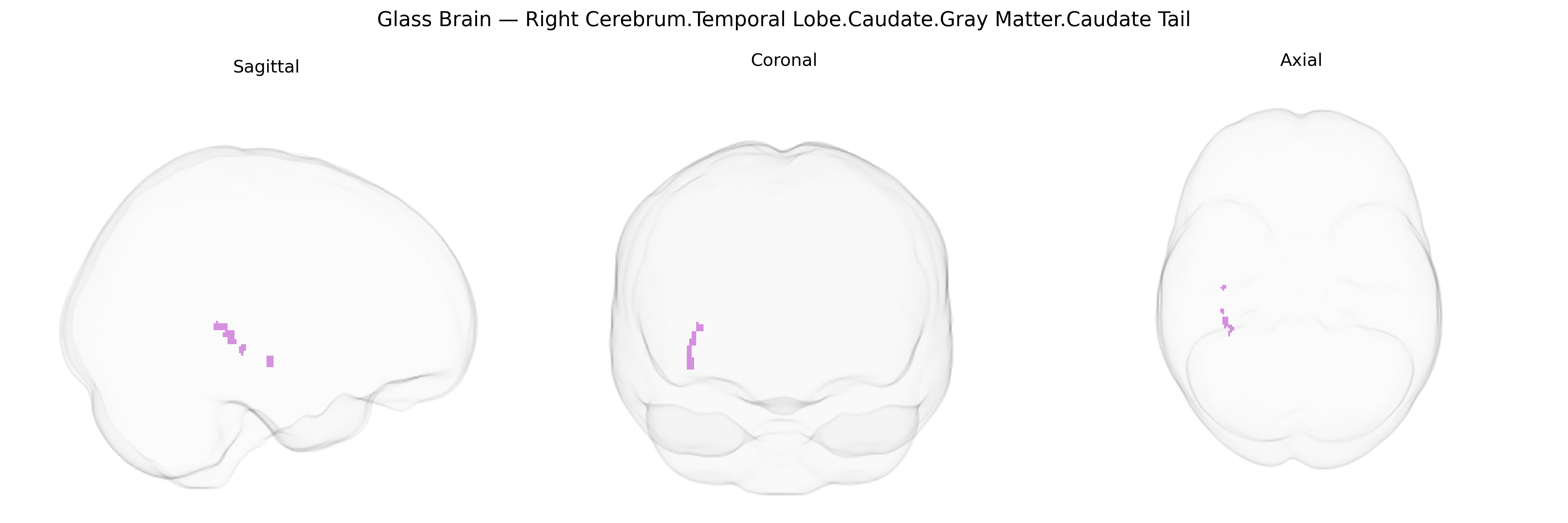
The bilateral Right Cerebrum.Temporal Lobe.Caudate.Gray Matter.Caudate Tail region corresponds to the posterior portion of the caudate nucleus, a C-shaped gray matter structure of the dorsal striatum that arches posteriorly into the temporal lobe along the lateral ventricle. The caudate tail participates in cortico-striato-thalamo-cortical loops, receiving inputs from association cortices (including temporal regions involved in visual and mnemonic processing) and projecting via basal ganglia output pathways to influence thalamic and cortical activity. Functionally, this segment of the caudate has been implicated in higher-order sensorimotor integration, aspects of visual attention and recognition (particularly object and motion processing), and certain forms of habit learning and memory, reflecting its role in linking temporal cortical information with basal ganglia circuits for action selection and behavioral regulation. There is no direct link for the “caudate tail” as a standalone article; see the related structure Caudate nucleus.
The caudate tail, a posterior segment of the caudate nucleus extending into the temporal lobe, has been implicated in genetic studies of striatal volume, frontostriatal circuitry, and neuropsychiatric risk: large-scale GWAS of subcortical brain structures (e.g., ENIGMA and UK Biobank) have identified loci near genes such as PDE1C, FAT3, DCC, and NPTN associated with caudate volume or morphology, some of which may extend to tail-specific variation, although most imaging-genetics work aggregates the caudate as a whole rather than isolating the tail. Polygenic influences on caudate structure overlap with risk alleles for schizophrenia, bipolar disorder, ADHD, obsessive-compulsive disorder, and major depression, consistent with the caudate tail’s role in cortico-striato-thalamo-cortical loops and reward/associative learning; striatal genes affecting dopamine signaling (e.g., DRD2, DAT1/SLC6A3) and synaptic plasticity (e.g., BDNF) have shown associations with striatal volumes and functional responses that likely include tail involvement. In neurodegenerative disease, Huntington’s disease (HTT CAG expansions) and other monogenic basal ganglia disorders produce early and marked caudate atrophy that encompasses the tail, while GWAS of Parkinson’s disease and related movement disorders (involving genes such as GBA and LRRK2) implicate striatal circuits including the caudate tail in motor and cognitive symptoms. Additional GWAS have linked caudate structure and connectivity—again typically not tail-specific—to cognitive traits (general intelligence, working memory), educational attainment, and substance use phenotypes, with shared genetic architecture between caudate-associated variants and polygenic scores for these traits, suggesting that genetically modulated variability in caudate tail gray matter is part of broader networks influencing cognition, habit learning, and vulnerability to psychiatric and substance-use disorders.
Overview generated by GPT-4o (2026).
Region ID: 350
Hemisphere: bilateral
Atlas: Talairach labels 1mm
Full Quality Version: Download MP4
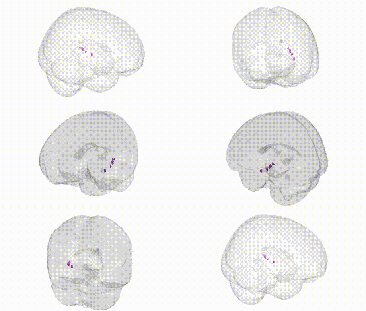
Full Quality Version: Download MP4
Wali Sidiqyar*, Gaurav Rudravaram*, Elyssa M. McMaster, Trent M. Schwartz, Adam M. Saunders, Kurt G. Schilling, Bennett A. Landman "Introducing SPINS: A Shared Public Visualization Library of Neuroanatomical Structures." Medical Imaging with Deep Learning- short paper
This resource is licensed under CC0 1.0 Universal (Public Domain).