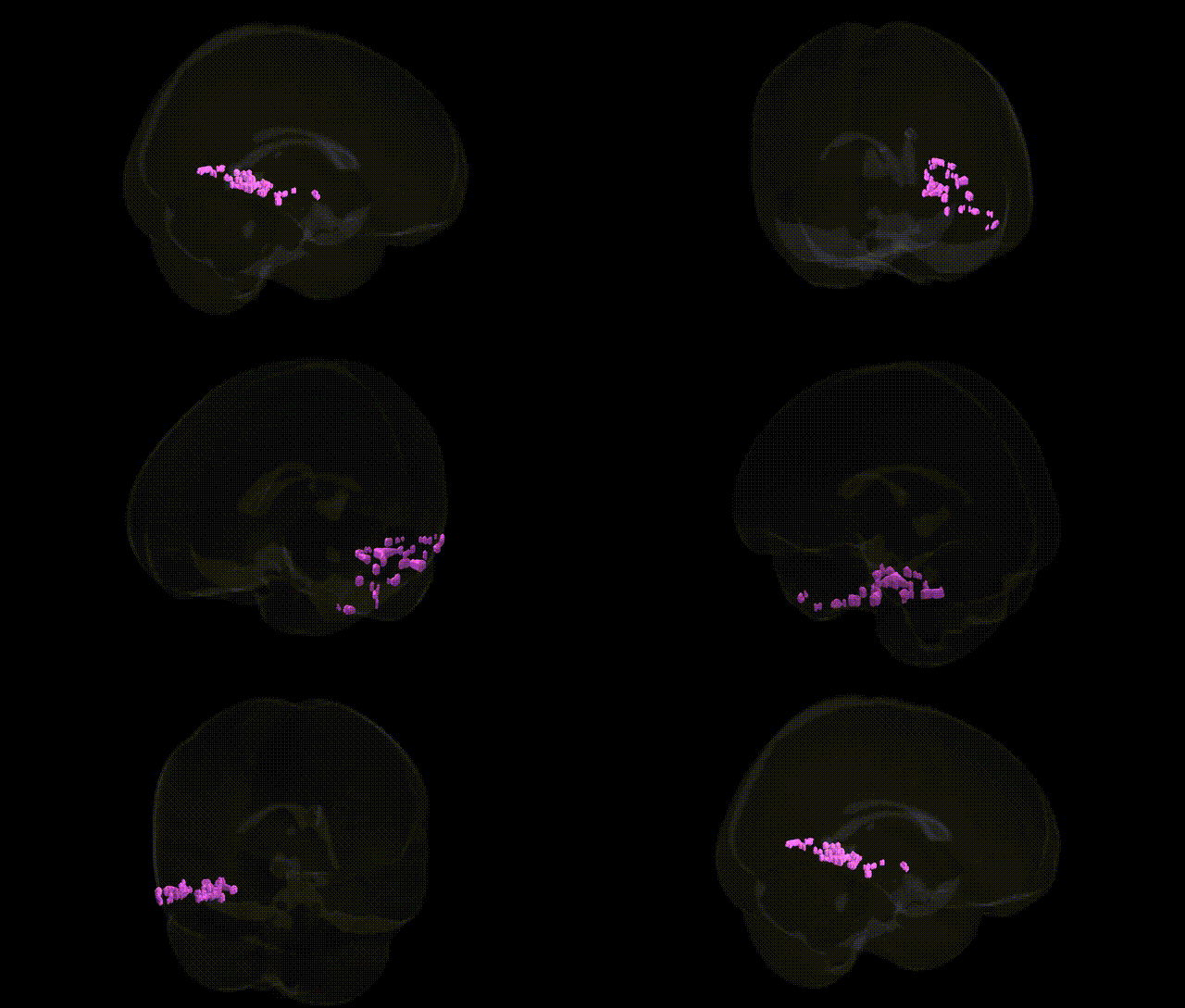
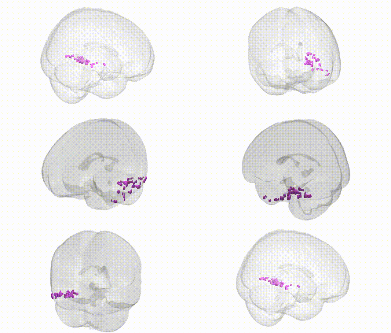
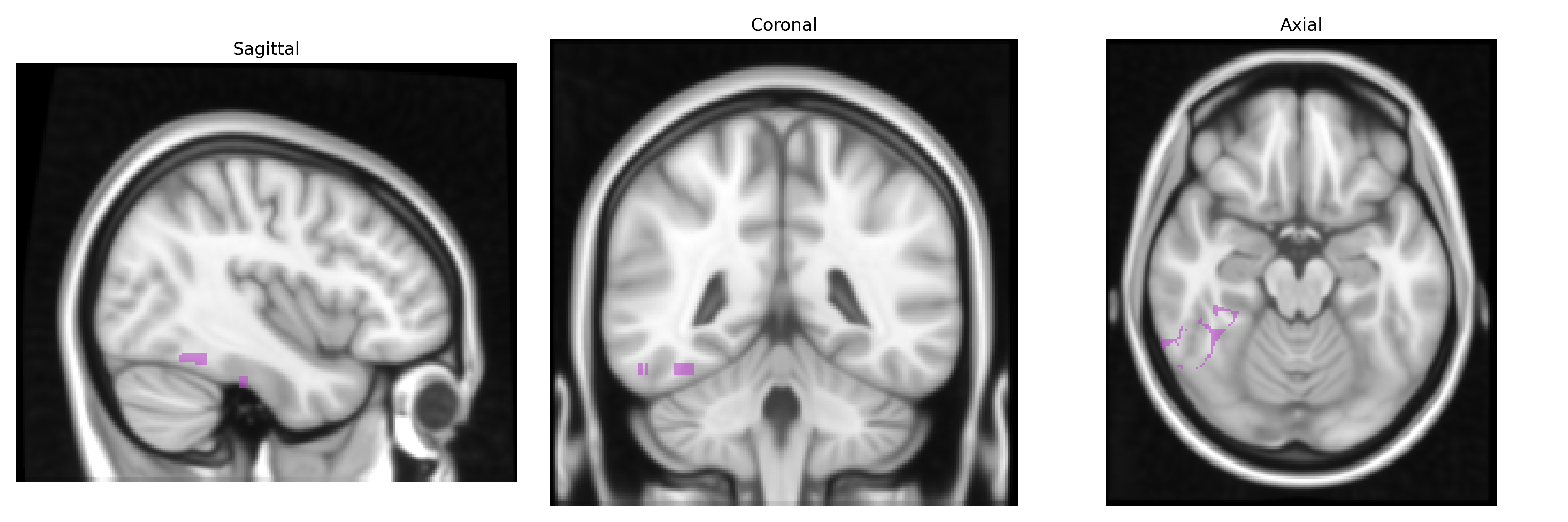
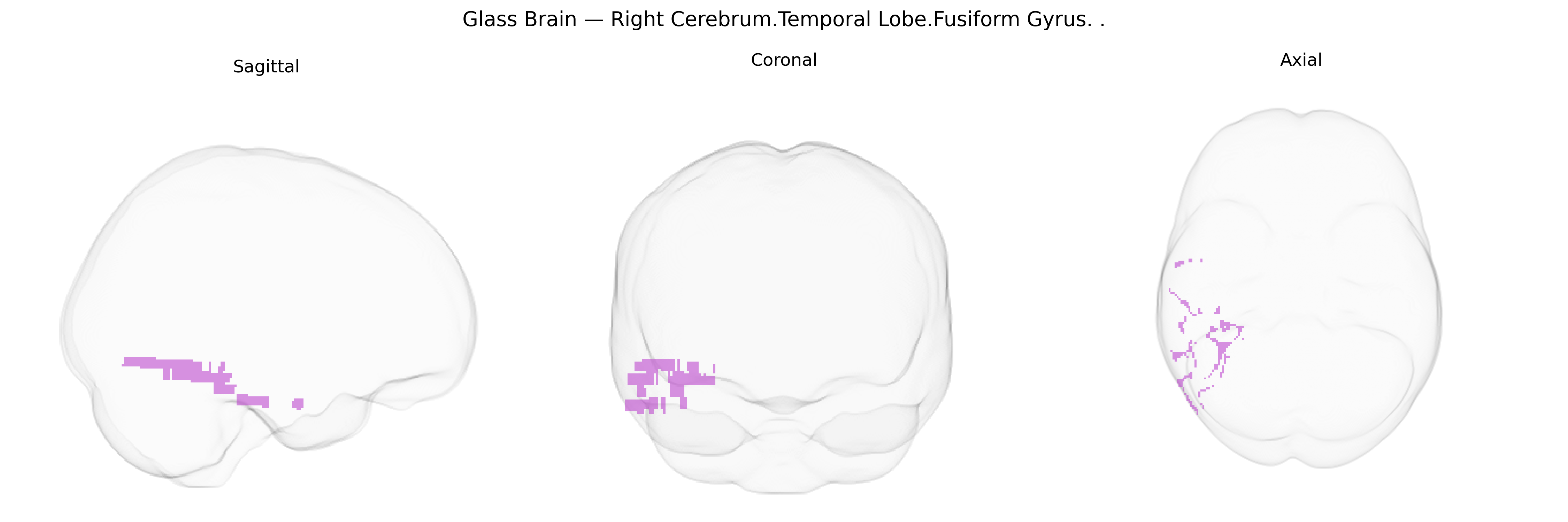
The bilateral Right Cerebrum.Temporal Lobe.Fusiform Gyrus, as defined in the Talairach 1 mm Atlas, corresponds to the fusiform gyrus located on the basal surface of the temporal and occipital lobes, extending longitudinally between the inferior temporal gyrus laterally and the parahippocampal gyrus medially. This region is involved in high-level visual processing, including object, face, and word recognition, and participates in category-specific representations (such as faces in the fusiform face area and visual word forms in the visual word form area). It receives input from earlier visual areas and interfaces with limbic and association cortices, supporting integration of visual features with memory and semantic information. Functionally, the fusiform gyrus shows hemispheric specialization patterns, with right-sided regions often more engaged in face and complex object perception, while still contributing bilaterally to a broad range of visual recognition tasks. Fusiform gyrus
The bilateral right fusiform gyrus of the temporal lobe has been repeatedly implicated in genetic studies of brain structure and cognition, with twin and heritability analyses showing substantial genetic control over fusiform volume and cortical thickness, often in the range of 40–70% heritability. GWAS of MRI-derived cortical measures (e.g., ENIGMA and UK Biobank consortia) have identified multiple loci associated with fusiform morphology, including variants near genes involved in neurodevelopment and synaptic function such as KIAA0586, DLG2, and others, although these are typically reported for “fusiform gyrus” bilaterally rather than specifically right-sided Talairach-defined parcels. Fusiform structure and function are genetically linked to face recognition ability, with rare variants and common polymorphisms in synaptic and neurodevelopmental genes contributing to individual differences in fusiform activation during face processing tasks, and to conditions such as developmental prosopagnosia. In autism spectrum disorder, polygenic risk and rare high-impact variants affecting genes such as NRXN1, SHANK3, and SCN2A have been associated with atypical fusiform activation and altered fusiform volume, particularly in social perception and face-processing paradigms. In schizophrenia and bipolar disorder, GWAS-derived polygenic risk scores correlate with fusiform structural alterations and connectivity changes, suggesting that shared genetic liability for psychosis influences temporal fusiform circuitry. Additional imaging genetics studies link fusiform measures to genetic risk for dyslexia, language and reading traits, and broader cognitive performance, indicating that fusiform morphology serves as an intermediate phenotype through which neurodevelopmental and psychiatric risk variants exert effects on higher-order visual and social cognition. While Talairach 1 mm labels provide anatomical precision, most genetic associations are reported at the level of the fusiform gyrus as a whole rather than specifically the bilateral right parcel, so current evidence supports a genetically influenced role for this region in face and object recognition, social cognition, and vulnerability to neurodevelopmental and psychiatric disorders without fine-grained lateralized genetic mapping.
Overview generated by GPT-4o (2026).
Region ID: 122
Hemisphere: bilateral
Atlas: Talairach labels 1mm
Full Quality Version: Download MP4
Full Quality Version: Download MP4
Wali Sidiqyar*, Gaurav Rudravaram*, Elyssa M. McMaster, Trent M. Schwartz, Adam M. Saunders, Kurt G. Schilling, Bennett A. Landman "Introducing SPINS: A Shared Public Visualization Library of Neuroanatomical Structures." Medical Imaging with Deep Learning- short paper
This resource is licensed under CC0 1.0 Universal (Public Domain).