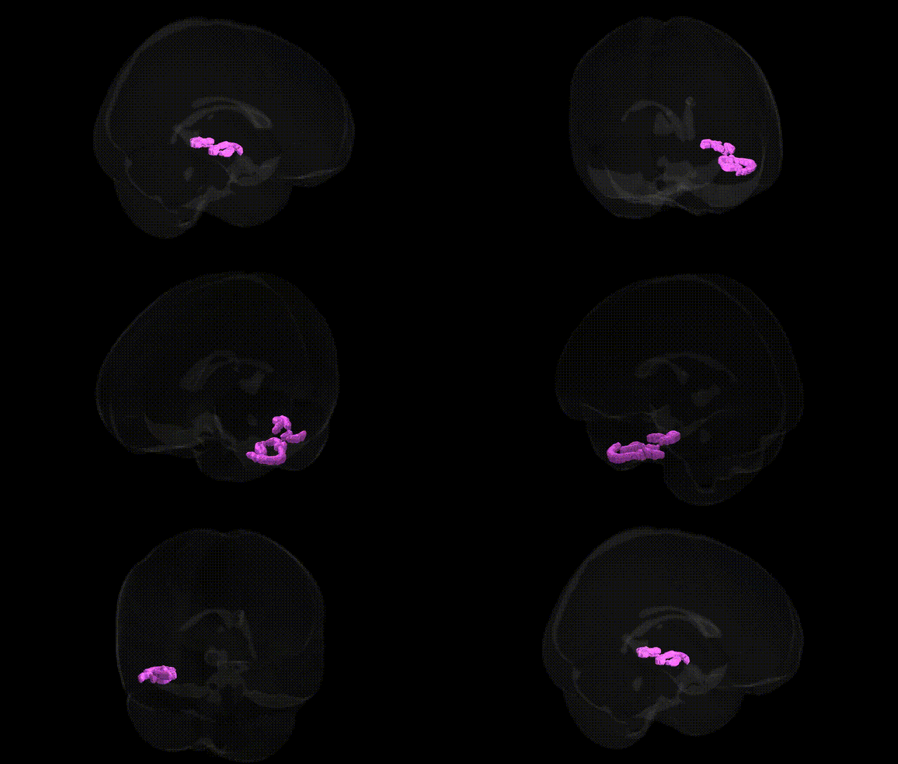
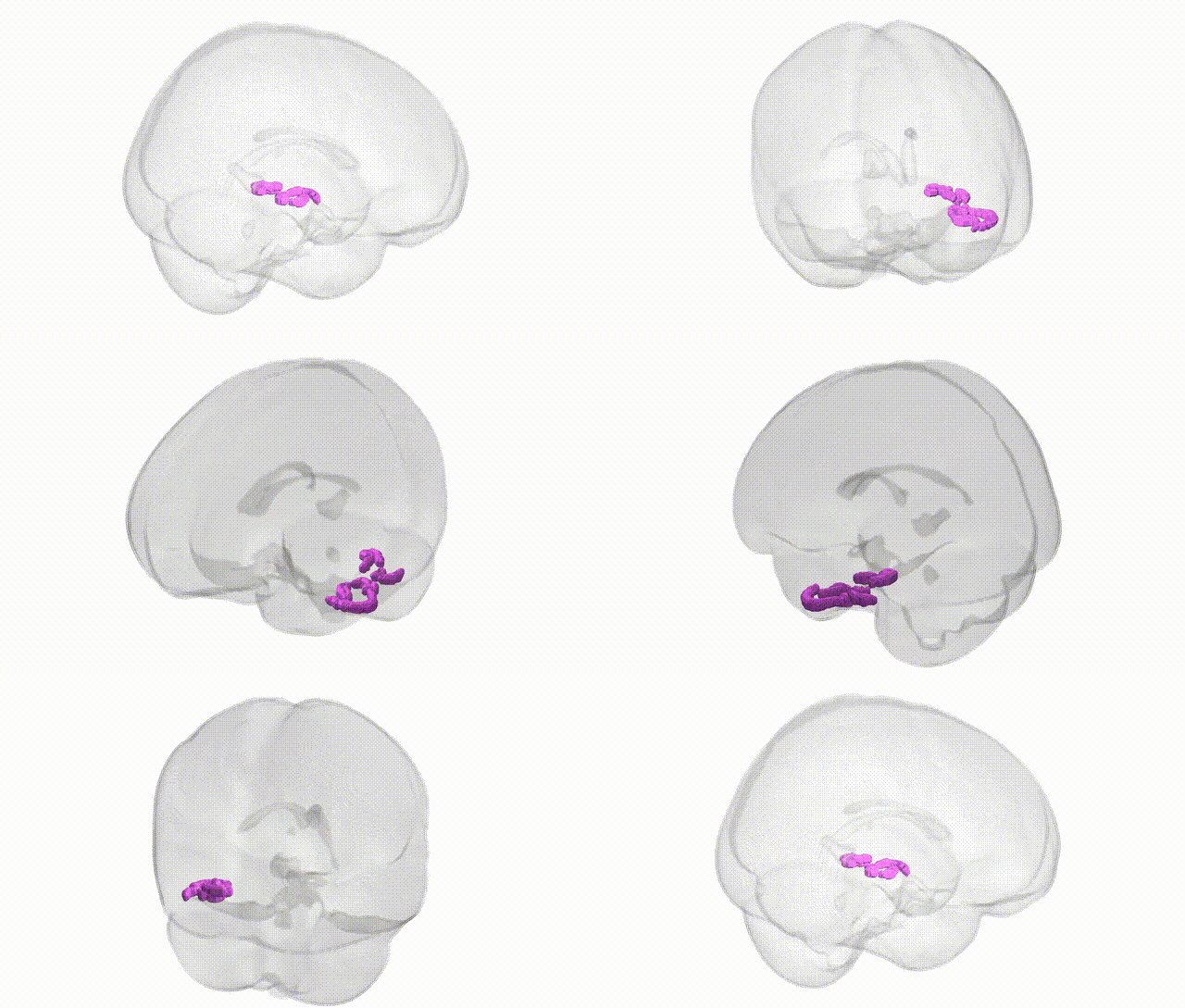
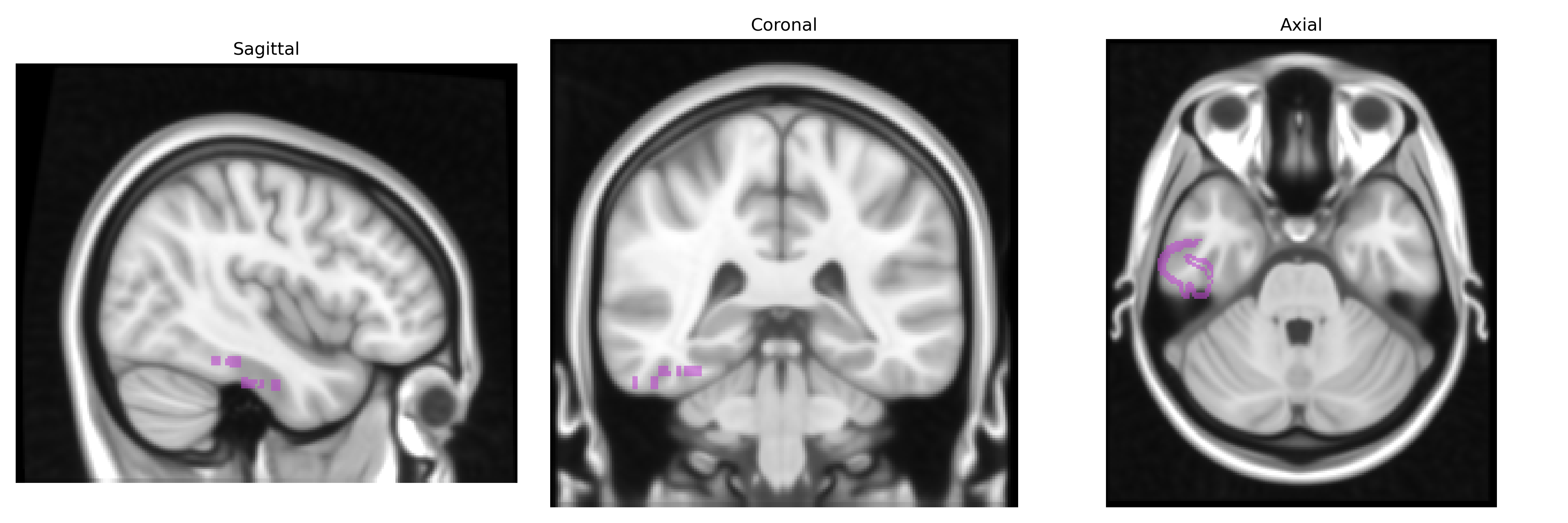
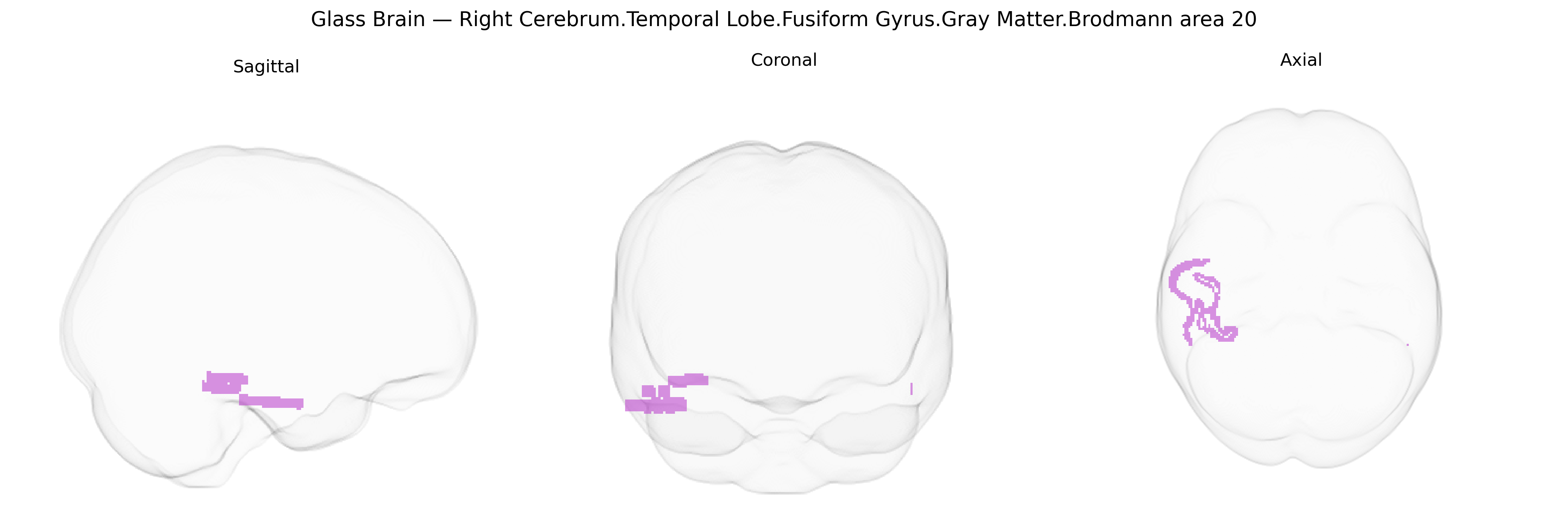
Bilateral right cerebrum temporal lobe fusiform gyrus gray matter Brodmann area 20 corresponds to a high-order visual association cortex located on the ventral surface of the temporal lobe, within the fusiform gyrus. This region participates in complex object recognition, including the processing of shapes, faces, and categories of visual stimuli, and it contributes to semantic integration of visual information with stored knowledge. BA20 receives input from primary and secondary visual areas and projects to distributed temporal and frontal networks involved in perception and memory, supporting functions such as visual identification and aspects of language related to object naming. There is no direct link for Brodmann area 20 in the fusiform gyrus; a closely related structure is the Fusiform gyrus.
The bilateral right fusiform gyrus gray matter in Brodmann area 20, as defined in the Talairach 1 mm atlas, has been implicated in several genetic and genome-wide association studies linking variability in this region to both structural and functional phenotypes. GWAS of cortical morphology (e.g., ENIGMA and UK Biobank–based studies) have identified common variants in genes involved in neurodevelopment, synaptic function, and cell adhesion—such as those near or within DCC, HMGA2, and various glutamatergic and neurodevelopmental loci—as associated with fusiform cortical thickness, surface area, or overall temporal lobe volume, although associations are often reported at the level of larger temporal or fusiform ROIs rather than precisely restricted BA20. Genetic studies of face processing and category-specific visual recognition highlight the fusiform “face area,” with heritability estimates indicating moderate to high genetic influence on fusiform activation patterns and structure; rare and common variants contributing to neurodevelopmental conditions such as autism spectrum disorder and congenital prosopagnosia have been linked to altered fusiform structure or function. Large GWAS of schizophrenia, bipolar disorder, and major depression show polygenic effects on temporal and fusiform volumes, reflecting shared genetic architecture between fusiform morphology and psychiatric risk loci, while Alzheimer’s disease and other neurodegenerative GWAS point to fusiform and inferior temporal atrophy patterns associated with risk variants in APOE and other amyloid- and tau-related genes. Collectively, current evidence indicates that fusiform BA20 structure and function, including in the right hemisphere, is a heritable trait influenced by widespread polygenic variation, with genetic associations converging on neurodevelopmental, synaptic, and neurodegenerative pathways that also underlie susceptibility to psychiatric and cognitive disorders.
Overview generated by GPT-4o (2026).
Region ID: 124
Hemisphere: bilateral
Atlas: Talairach labels 1mm
Full Quality Version: Download MP4
Full Quality Version: Download MP4
Wali Sidiqyar*, Gaurav Rudravaram*, Elyssa M. McMaster, Trent M. Schwartz, Adam M. Saunders, Kurt G. Schilling, Bennett A. Landman "Introducing SPINS: A Shared Public Visualization Library of Neuroanatomical Structures." Medical Imaging with Deep Learning- short paper
This resource is licensed under CC0 1.0 Universal (Public Domain).